. 2014 Apr 15;82(15):1331-8.
doi: 10.1212/WNL.0000000000000312. Epub 2014 Mar 12.

Vascular risk factors, large-artery atheroma, and brain white matter hyperintensities

Affiliations

Vascular risk factors, large-artery atheroma, and brain white matter hyperintensities

Joanna M Wardlaw et al. Neurology. .

Abstract

Objective: To determine the magnitude of potentially causal relationships among vascular risk factors (VRFs), large-artery atheromatous disease (LAD), and cerebral white matter hyperintensities (WMH) in 2 prospective cohorts.

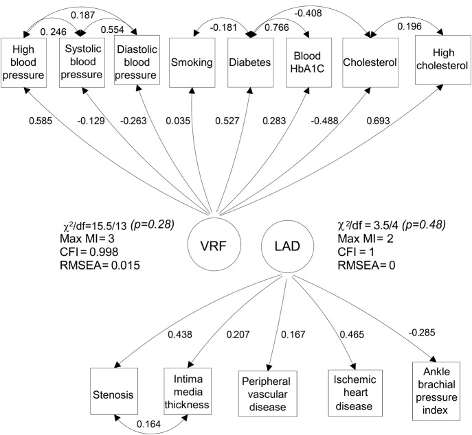
Methods: We assessed VRFs (history and measured variables), LAD (in carotid, coronary, and leg arteries), and WMH (on structural MRI, visual scores and volume) in: (a) community-dwelling older subjects of the Lothian Birth Cohort 1936, and (b) patients with recent nondisabling stroke. We analyzed correlations, developed structural equation models, and performed mediation analysis to test interrelationships among VRFs, LAD, and WMH.

Results: In subjects of the Lothian Birth Cohort 1936 (n = 881, mean age 72.5 years [SD ±0.7 years], 49% with hypertension, 33% with moderate/severe WMH), VRFs explained 70% of the LAD variance but only 1.4% to 2% of WMH variance, of which hypertension explained the most. In stroke patients (n = 257, mean age 74 years [SD ±11.6 years], 61% hypertensive, 43% moderate/severe WMH), VRFs explained only 0.1% of WMH variance. There was no direct association between LAD and WMH in either sample. The results were the same for all WMH measures used.

Conclusions: The small effect of VRFs and LAD on WMH suggests that WMH have a large "nonvascular," nonatheromatous etiology. VRF modification, although important, may be limited in preventing WMH and their stroke and dementia consequences. Investigation of, and interventions against, other suspected small-vessel disease mechanisms should be addressed.


Figures

Figure 1
Figure 1. LBC1936 cohort: Diagram of measurement models for the VRF and LAD constructs
Standardized loadings and residual correlations are shown. Numbers adjacent to paths may be squared to obtain the shared variance between adjacent variables. Double-headed arrows are correlations; single-headed arrows are hypothesized causal pathways. The convention used represents manifest (measured) variables as rectangles and constructed variables (VRFs or LAD) as circles. Model fit parameters are shown adjacent to the constructed variable: a nonsignificant χ2 is a sign of a well-fitting model; the Max MI (which indicates the degree of greatest local strain within the model in terms of a potential reduction in model χ2); the CFI (≥0.90 indicates acceptable fit); and the RMSEA (<0.06 indicates acceptable fit). CFI = comparative fit index; HbA1c = hemoglobin A1c; LAD = large-artery atheromatous disease; LBC1936 = Lothian Birth Cohort 1936; Max MI = maximum modification index; RMSEA = root mean square error of approximation; VRF = vascular risk factor.
Figure 2
Figure 2. Structural models in the LBC1936
(A) This model is the total association between LAD and WMH. (B) Hierarchical association with VRFs controlled. (C) Total effect of VRFs on WMH. (D) The mediation model (see the text). Standardized regression coefficients (parameter weights) are shown adjacent to each path. Each arrow is directed from a predictor variable to the outcome variable. In model B, the 0.828 indicates that approximately 70% of the variance in LAD is explained by VRFs and the 0.111 indicates that approximately 2% of the variance in WMH is explained by VRFs. The 0.292 and 0.984 adjacent to the lateral arrows in model B indicate the variance that is unexplained by VRFs on LAD and WMH, respectively. The estimates shown in the figure are for WMH measured using combined periventricular and deep Fazekas scores. The estimates for the model using other WMH measures are shown in table 2. R2 is the WMH model R2 value; s2 is the residual variance. LAD = large-artery atheromatous disease; LBC1936 = Lothian Birth Cohort 1936; VRF = vascular risk factor; WMH = white matter hyperintensity.

References

    1. Debette S, Markus HS. The clinical importance of white matter hyperintensities on brain magnetic resonance imaging: systematic review and meta-analysis. BMJ 2010;341:c3666. - PMC - PubMed
    1. Baezner H, Blahak C, Poggesi A, et al. Association of gait and balance disorders with age-related white matter changes: the LADIS study. Neurology 2008;70:935–942 - PubMed
    1. Wardlaw JM, Smith C, Dichgans M. Mechanisms of sporadic cerebral small vessel disease: insights from neuroimaging. Lancet Neurol 2013;12:483–497 - PMC - PubMed
    1. Marcus J, Gardener H, Rundek T, et al. Baseline and longitudinal increases in diastolic blood pressure are associated with greater white matter hyperintensity volume: the Northern Manhattan Study. Stroke 2011;42:2639–2641 - PMC - PubMed
    1. Guo X, Pantoni L, Simoni M, et al. Blood pressure components and changes in relation to white matter lesions: a 32-year prospective population study. Hypertension 2009;54:57–62 - PubMed

Publication types