

Wound morphology and topography in the diabetic foot: hurdles in implementing angiosome-guided revascularization
- PMID: 24624299
- PMCID: PMC3929512
- DOI: 10.1155/2014/672897
Wound morphology and topography in the diabetic foot: hurdles in implementing angiosome-guided revascularization
Abstract
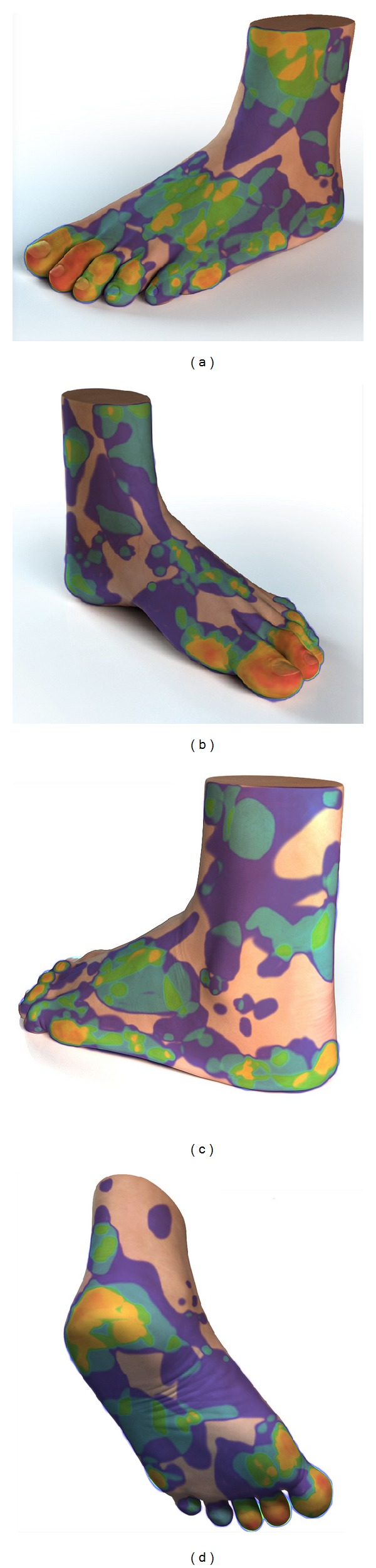
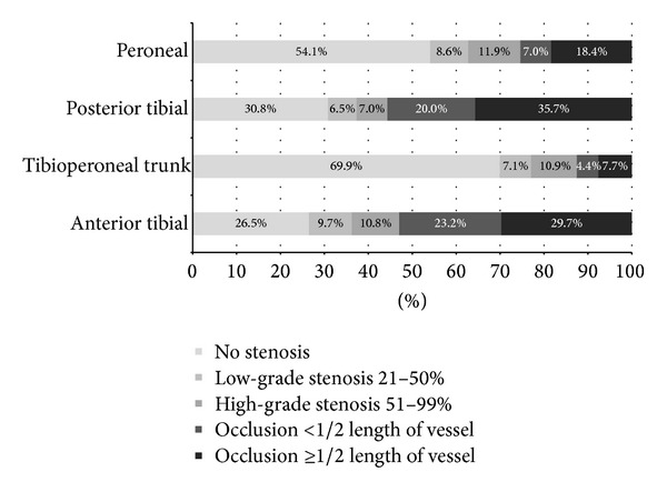
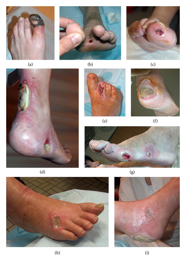
Purpose. Angiosome-guided revascularization is an approach that improves wound healing but requires a surgeon to determine which angiosomes are ischemic. This process can be more difficult than anticipated because diabetic foot (DF) wounds vary greatly in quantity, morphology, and topography. This paper explores to what extent the heterogeneous presentation of DF wounds impedes development of a proper revascularization strategy. Methods. Data was retrieved from a registry of patients scheduled for below-the-knee (BTK) revascularization. Photographs of the foot and historic benchmark diagrams were used to assign wounds to their respective angiosomes. Results. In 185 limbs we detected 345 wounds. Toe wounds (53.9%) could not be designated to a specific angiosome due to dual blood supply. Ambiguity in wound stratification into angiosomes was highest at the heel, achilles tendon, and lateral/medial side of the foot and lowest for malleolar wounds. In 18.4% of the DF, at least some wounds could not confidently be categorized. Proximal wounds (coinciding with toe wounds) further steered revascularization strategy in 63.6%. Multiple wounds required multiple BTK revascularization in 8.6%. Conclusion. The heterogeneous presentation in diabetic foot wounds hampers unambiguous identification of ischemic angiosomes, and as such diminishes the capacity of the angiosome model to optimize revascularization strategy.
Figures
References
-
- Faglia E, Mantero M, Caminiti M, et al. Extensive use of peripheral angioplasty, particularly infrapopliteal, in the treatment of ischaemic diabetic foot ulcers: clinical results of a multicentric study of 221 consecutive diabetic subjects. Journal of Internal Medicine. 2002;252(3):225–232. - PubMed
-
- Neville RF, Attinger CE, Bulan EJ, Ducic I, Thomassen M, Sidawy AN. Revascularization of a specific angiosome for limb salvage: does the target artery matter? Annals of Vascular Surgery. 2009;23(3):367–373. - PubMed
-
- Alexandrescu V-A, Hubermont G, Philips Y, et al. Selective primary angioplasty following an angiosome model of reperfusion in the treatment of Wagner 1-4 diabetic foot lesions: practice in a multidisciplinary diabetic limb service. Journal of Endovascular Therapy. 2008;15(5):580–593. - PubMed
-
- Alexandrescu V, Söderström M, Venermo M. Angiosome theory: fact or fiction? Scandinavian Journal of Surgery. 2012;101(2):125–131. - PubMed
-
- Azuma N, Uchida H, Kokubo T, Koya A, Akasaka N, Sasajima T. Factors influencing wound healing of critical ischaemic foot after bypass surgery: is the angiosome important in selecting bypass target artery? European Journal of Vascular and Endovascular Surgery. 2012;43(3):322–328. - PubMed
LinkOut - more resources
Full Text Sources
Other Literature Sources
Miscellaneous
