

Self-assembly of large-scale gold nanoparticle arrays and their application in SERS
- PMID: 24624899
- PMCID: PMC3995606
- DOI: 10.1186/1556-276X-9-114
Self-assembly of large-scale gold nanoparticle arrays and their application in SERS
Abstract
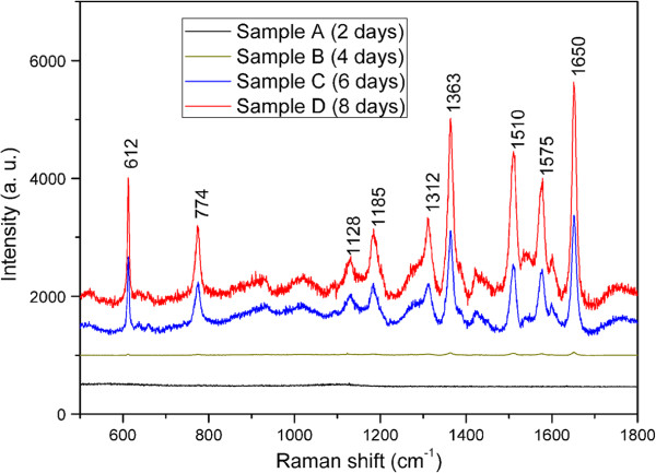
Surface-enhanced Raman scattering is an effective analytical method that has been intensively applied in the field of identification of organic molecules from Raman spectra at very low concentrations. The Raman signal enhancement that makes this method attractive is usually ascribed to the noble metal nanoparticle (NMNP) arrays which can extremely amplify the electromagnetic field near NMNP surface when localized surface plasmon resonance (LSPR) mode is excited. In this work, we report a simple, facile, and room-temperature method to fabricate large-scale, uniform gold nanoparticle (GNP) arrays on ITO/glass as SERS substrates using a promoted self-assembly deposition technique. The results show that the deposition density of GNPs on ITO/glass surface increases with prolonging deposition time, and nanochain-like aggregates appear for a relatively longer deposition time. It is also shown that these films with relatively higher deposition density have tremendous potential for wideband absorption in the visible range and exhibit two LSPR peaks in the extinction spectra because the electrons simultaneously oscillate along the nanochain at the transverse and the longitudinal directions. The SERS enhancement activity of these GNP arrays was determined using 10-6 M Rhodamine 6G as the Raman probe molecules. A SERS enhancement factor as large as approximately 6.76 × 10⁶ can be obtained at 1,363 cm-1 Raman shift for the highest deposition density film due to the strong plasmon coupling effect between neighboring particles.
Figures


References
LinkOut - more resources
Full Text Sources
Other Literature Sources
Miscellaneous
