Review
. 2014 Apr;14(4):449-63.
doi: 10.1586/14737175.2014.896199.

Applications of SPR for the characterization of molecules important in the pathogenesis and treatment of neurodegenerative diseases

Affiliations
Review

Applications of SPR for the characterization of molecules important in the pathogenesis and treatment of neurodegenerative diseases

Nathan J Wittenberg et al. Expert Rev Neurother. 2014 Apr.

Abstract

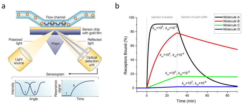
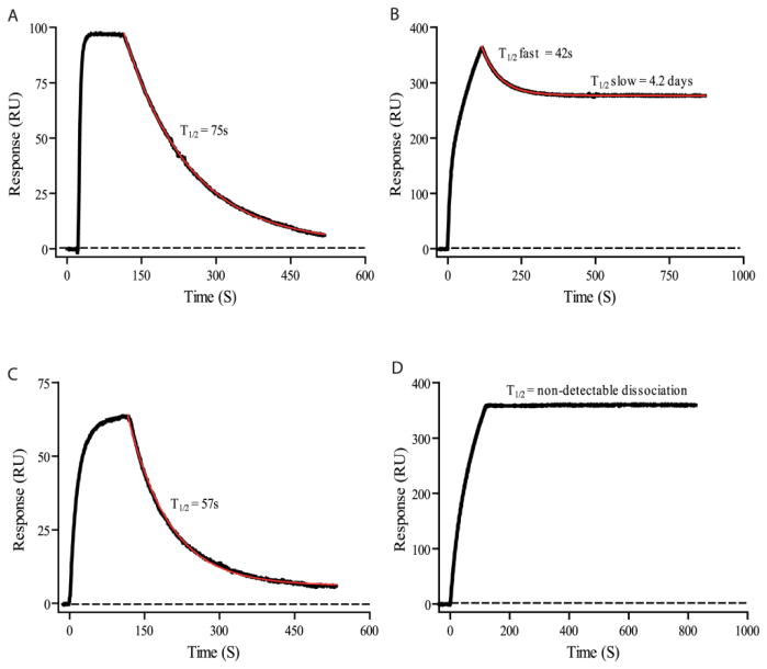
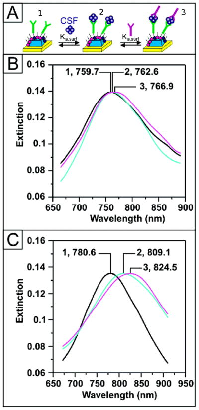
Characterization of binding kinetics and affinity between a potential drug and its receptor are key steps in the development of new drugs. Among the techniques available to determine binding affinities, surface plasmon resonance has emerged as the gold standard because it can measure binding and dissociation rates in real-time in a label-free fashion. Surface plasmon resonance is now finding applications in the characterization of molecules for treatment of neurodegenerative diseases, characterization of molecules associated with pathogenesis of neurodegenerative diseases and detection of neurodegenerative disease biomarkers. In addition it has been used in the characterization of a new class of natural autoantibodies that have therapeutic potential in a number of neurologic diseases. In this review we will introduce surface plasmon resonance and describe some applications of the technique that pertain to neurodegenerative disorders and their treatment.


Figures

Figure 1
Figure 1
(a) Schematic of typical SPR device. Light is directed onto a gold film and the reflection is monitored with a detector. At a certain angle, the light does not reflect but generates surface plasmon resonance (SPR). In a flow cell above the film, receptors are linked to the film and analyte solution is flowed past the receptors. If the analyte binds to the receptors, the angle at which SPR is generated shifts, and this angle is tracked over time as the reaction progresses. (b) Simulated SPR binding curves of four different molecules (A,B,C,D) with identical KD values (10 nM), but different rate constants, kon and koff. Analyte concentration was 100 nM for all molecules. Units for kon are M−1s−1 and for koff, s−1. This plot demonstrates the significance of knowing the kinetic constants. Molecule A would bind quickly and dissociate quickly, whereas molecule D would bind slowly but dissociate very slowly. The half-lives (t1/2 = ln 2/koff) of the bound complexes are roughly (from A–D): 12 min, 2 hr, 20 hr, and 8 days. (a) is adapted from Cooper [9] and used with permission of Macmillan Publishers Ltd.
Figure 2
Figure 2
Immobilization of a receptor (R) with a primary amine on a self assembled monolayer (SAM) via EDC/NHS coupling. The carboxyl-terminated thiol is converted to a succinimide ester by reaction with carbodiimide (EDC) and N-hydroxylsuccinimide (NHS), making it reactive toward molecules with available primary amines, as shown by the circle marked “R”. The final product is a receptor (R) covalently linked to the surface with an amide bond.
Figure 3
Figure 3
SPR characterization of remyelinating IgMs binding to supported lipid bilayers (SLB). (a) SPR kinetics curves of antibody O4 (5 – 50 nM) binding to a SLB containing 2 mole % sulfatide (Sulf). (b) SPR kinetics curves of antibody O1 (5 – 50 nM) binding to a SLB containing 2 mole % galactocerebroside (GalC). (c) Negative controls showing that neither O1 nor O4 bind to SLBs composed solely of egg phosphatidylcholine (egg PC) (solid lines). O1 and O4 did not bind to the sensor protected only by BSA blocking in the absence of a SLB (dashed lines). (d) SPR kinetics curves for 1.25 nM and 310 pM O4 binding to a SLB that mimics the Sulf and GalC content of myelin: 6 mole % Sulf, 16 mole % GalC. In all panels the solid black lines are exponential fits to the data used to extract association and dissociation rate constants. From Wittenberg et al., [67] and used with permission of the American Chemical Society.
Figure 4
Figure 4
High-affinity monoclonal, recombinant anti–aquaporin-4 (AQP4) antibody (rAb) for antibody blocking therapy. (a) Crystal structure of AQP4 tetramer is shown on the same scale with that of an IgG antibody. (b) SPR measurements of recombinant antibody rAb-53 binding to AQP4-reconstituted proteoliposomes. Antibody rAb-53 ranged in concentration from 0.13 to 2.2 μM. (c) SPR measurements of additional recombinant antibodies binding to AQP4 in proteoliposomes. (d) SPR measurements of rAb-53 with the L243A/L235A mutations binding to AQP4 proteoliposomes. These mutations eliminated complement-dependent cytotoxicity and antibody-dependent cell-mediated cytotoxicity, but did not significantly alter the binding properties. Adapted from Tradtrantip et al., [18] and used with permission of John Wiley and Sons.
Figure 5
Figure 5
SPR characterization of binding of a monoclonal IgM, OMAB, to Aβ peptide monomers and oligomers. OMAB was immobilized on the chip followed by injections of (A) Aβ(1-40) monomers or (B) oligomers, or (C) Aβ(1-42) monomers or (D) oligomers. The dissociation curves show that monomers do not stay bound to the IgM very long (t1/2 = ~1 min), whereas the oligomers stay bound to the IgM for much longer (t1/2 = days, or longer), indicating preference for the toxic oligomers. Thus, these IgMs may help capture oligomers for immune system processing, or be used as a capture molecule in a detection scheme. Figure is from Lindhagen-Persson et al. [103]
Figure 6
Figure 6
Detection of Alzheimer’s disease biomarker ADDL in human CSF samples using a sandwich assay and SPR. The SPR signal is monitored as peaks in the extinction (i.e. absorption) spectrum of the SPR sensor and molecular binding is indicated by a red-shift in the spectrum. (A) Illustration showing (1) anti-ADDL immobilization on the SPR sensor, (2) capture of ADDL, and (3) addition of second anti-ADDL to amplify changes is SPR signal. (B) Sensor response to CSF of an aging control patient: After functionalization with (B-1) 100 nM anti-ADDL (λmax = 759.7 nm), (B-2) CSF (λmax= 762.6 nm), and (B-3) 100 nM anti-ADDL (λmax = 766.9 nm). (C) Sensor response to CSF from an AD patient: After functionalization with (C-1) 100 nM anti-ADDL (λmax = 780.6 nm), (C-2) CSF (λmax = 809.1 nm), and (C-3) 100 nM anti-ADDL (λmax = 824.5 nm). Figure adapted from Haes et al.[108] and used with permission of the American Chemical Society.

References

    1. Arvanitakis Z, Grodstein F, Bienias JL, et al. Relation of NSAIDs to incident AD, change in cognitive function, and AD pathology. Neurology. 2008;70(23):2219–2225. - PubMed
    1. Driver JA, Logroscino G, Lu L, Gaziano JM, Kurth T. Use of non-steroidal anti-inflammatory drugs and risk of Parkinson’s disease: nested case-control study. BMJ. 2011;342:d198. - PMC - PubMed
    1. Martin BK, Szekely C, Brandt J, et al. Cognitive function over time in the Alzheimer’s Disease Anti-inflammatory Prevention Trial (ADAPT): results of a randomized, controlled trial of naproxen and celecoxib. Arch Neurol. 2008;65(7):896–905. - PMC - PubMed
    1. Consortium WTCC. Genome-wide association study of 14,000 cases of seven common diseases and 3,000 shared controls. Nature. 2007;447(7145):661–678. - PMC - PubMed
    1. Mizushima N, Levine B, Cuervo AM, Klionsky DJ. Autophagy fights disease through cellular self-digestion. Nature. 2008;451(7182):1069–1075. - PMC - PubMed

Publication types