. 2014 Mar 14;7(1):19.
doi: 10.1186/1757-1146-7-19.

Measurement of three-dimensional morphological characteristics of the calcaneus using CT image post-processing

Affiliations

Measurement of three-dimensional morphological characteristics of the calcaneus using CT image post-processing

Minfei Qiang et al. J Foot Ankle Res. .

Abstract

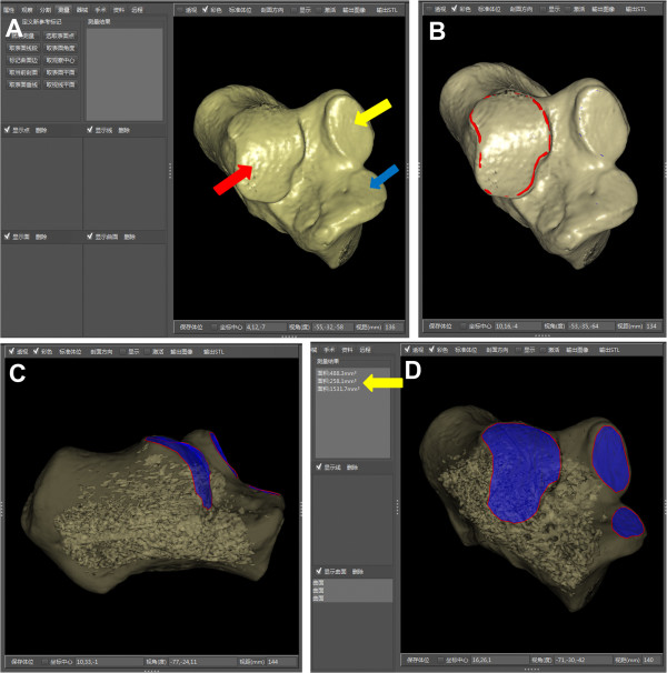
Background: Although computed tomography (CT) with three-dimensional (3D) rendering of the calcaneus is used for diagnostic evaluation of disorders, morphological measurements for the calcaneus are mostly based on a two-dimensional plane. The purposes of this study were to design a method for 3D morphological measurements of the normal calcaneus based on CT post-processing techniques, to measure morphological parameters in the male and female groups and describe gender differences of the parameters, and to investigate the reliability of such measurements.

Methods: One hundred and seventy-nine patients (83 men and 96 women) with a mean age of 40.6 (range, 21 to 59) years who underwent CT scans for their feet were reviewed retrospectively. The 3D structure of a normal calcaneus after shaded surface display reconstruction was extracted by interactive and automatic segmentation. Morphological measurements were achieved by means of a 3D measurement method based on CT image post-processing. Lengths and heights of the main parts of the calcaneus, Gissane's angle, Böhler's angle and the area of articular facet were worked out in 3D space. Gender-related size differences of parameters were compared using analysis of covariance (ANCOVA), adjusting for body height. Intra-observer and inter-observer reliabilities were assessed using intraclass correlation coefficients (ICCs) and the root mean square standard deviation (RMS-SD) for precision study.

Results: A large range of measurement values was found. Only the length of the anterior process was without gender difference (p > 0.05). The other parameters in the male group were greater than those in the female group (p < 0.01 for each, ANCOVA). All parameters had excellent reliability and reproducibility (ICC > 0.8). Precision was acceptable for intra-observer RMS-SD (linear, angular and areal measurements no more than 0.6 mm, 1.2° and 0.25 mm2, respectively). Inter-observer RMS-SD ranged from 0.4 to 1.6 mm for linear measurements, 1.2 to 2.5° for angles and 0.24 to 0.40 mm2 for areas.

Conclusions: Three-dimensional morphological measurement based on a CT post-processing technique was highly reliable and repeatable for calcaneal anatomic morphological measurement. The current data will be helpful for anatomic reduction of calcaneal fractures and calcaneal malunion.


Figures

Figure 1
Figure 1
Process of generating the 3D structures of the calcaneus. (A) Joints of the hindfoot containing the subtalar joint, calcaneocuboid joint and talonavicular joint combined with each other, which showed the blue clumps. These bones couldn’t be recognized automatically by surface rendering in 3D Imaging. (B) The calcaneus and other bones were labeled separately by 3D interactive and automatic segmentation. After the Enter key was clicked to start the computer processing, the color of labeled bones gradually turned red and green and then the division was finished automatically in one minute. (C) The calcaneus was marked red and others were marked green at last. (D) The calcaneus was extracted and other bones were deleted.
Figure 2
Figure 2
Three-dimensional morphological measurements of calcaneus. (A, B) Location of measurement points was illustrated. A = highest point of posterior facet; B = bottom of the posterior facet at lateral surface intersecting anterior process of calcaneus; C = highest point of calcaneocuboid joint; D = lowest point of calcaneocuboid joint; E = most posterior point of calcaneal tuberosity; F = highest point on the superior edge of calcaneal tuberosity. (C, D) The calcaneal morphological parameters in 3D space were shown. GE = length of calcaneal axis; AH = height of posterior facet; AB = length of posterior facet; BC = length of anterior process; CD = height of anterior process; ∠ABC = Gissane’s angle; 180°-∠CAF = Böhler’s angle. The posterior and middle facets were shown as blue surfaces.
Figure 3
Figure 3
Areal measurements of subtalar articular facets for example. (A) After the segmentation, the SSD reconstruction image of the 3D calcaneus presented the anterior (blue arrow), middle (yellow arrow) and posterior (red arrow) facets clearly. (B) The operator marked the border of the articular facet. The border (red curve) was actually unbroken, though part of the red curve seemed not visualized. (C) With the help of the perspective mode of SSD reconstruction, the defined boundary of the surface could be observed. (D) On the anterolateral view, the boundary of each articular facet was clear. Each area was calculated by the software (yellow arrow).
Figure 4
Figure 4
Intra-observer and inter-observer reliabilities of 3D morphological measurements (n = 36). The average ICC of inter-observer reliability was calculated from all obtained measurements among three examiners. All the parameters exhibited high ICC values, including length of calcaneal axis (LCA), height of posterior facet (HPF), length of posterior facet (LPF), length of anterior process (LAP), height of anterior process (HAP), Gissane's angle (GA), Böhler's angle (BA), area of posterior facet (APF), and area of middle facet (AMF).

References

    1. ESSEX-LOPRESTI P. The mechanism, reduction technique, and results in fractures of the os calcis. Br J Surg. 1952;39:395–419. doi: 10.1002/bjs.18003915704. - DOI - PubMed
    1. Potter MQ, Nunley JA. Long-term functional outcomes after operative treatment for intra-articular fractures of the calcaneus. J Bone Joint Surg Am. 2009;91:1854–1860. doi: 10.2106/JBJS.H.01475. - DOI - PubMed
    1. Sanders R, Fortin P, DiPasquale T, Walling A. Operative treatment in 120 displaced intraarticular calcaneal fractures. Results using a prognostic computed tomography scan classification. Clin Orthop Relat Res. 1993;290:87–95. - PubMed
    1. Mulcahy DM, McCormack DM, Stephens MM. Intra-articular calcaneal fractures: effect of open reduction and internal fixation on the contact characteristics of the subtalar joint. Foot Ankle Int. 1998;19:842–848. doi: 10.1177/107110079801901209. - DOI - PubMed
    1. Paley D, Hall H. Intra-articular fractures of the calcaneus. A critical analysis of results and prognostic factors. J Bone Joint Surg Am. 1993;75:342–354. - PubMed