Mixed epithelial and mesenchymal metaplastic carcinoma (carcinosarcoma) of the breast: a case report
- PMID: 24625319
- PMCID: PMC3976087
- DOI: 10.1186/2047-783X-19-14
Mixed epithelial and mesenchymal metaplastic carcinoma (carcinosarcoma) of the breast: a case report
Abstract
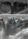
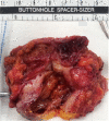
Metaplastic breast carcinoma (MBC) is an uncommon malignancy characterized by the co-existence of two or more cellular types, commonly a mixture of epithelial and mesenchymal components. A case of a female patient aged 46 years with MBC (carcinosarcoma) is presented, including mammographic, ultrasonic, gross examination, and pathological findings. After undergoing modified radical mastectomy of the left breast and subsequent six courses of adjuvant chemotherapy and endocrine therapy, the patient is now doing well with no recurrence and metastasis. Conventional treatments for invasive ductal carcinoma (IDC) may appear to be less effective. Patients with MBC would be appropriate candidates for innovative or targeted therapy regimens.
Figures


Similar articles
-
[Carcinosarcoma of the breast: a tumour with controversial histogenesis].Clin Transl Oncol. 2005 Jul;7(6):255-7. doi: 10.1007/BF02710172. Clin Transl Oncol. 2005. PMID: 16131449 Spanish.
-
Carcinosarcoma of the breast: report of two cases.Eur J Gynaecol Oncol. 2003;24(1):93-5. Eur J Gynaecol Oncol. 2003. PMID: 12691330
-
Metaplastic carcinoma of the breast with mesenchymal differentiation (carcinosarcoma). A unique presentation of an aggressive malignancy and literature review.Breast Dis. 2018;37(3):169-175. doi: 10.3233/BD-170313. Breast Dis. 2018. PMID: 29504519 Review.
-
Carcinosarcoma of the breast.J Coll Physicians Surg Pak. 2012 May;22(5):333-4. J Coll Physicians Surg Pak. 2012. PMID: 22538044
-
Metaplastic breast carcinoma: case series and review of the literature.Asian Pac J Cancer Prev. 2012;13(9):4645-9. doi: 10.7314/apjcp.2012.13.9.4645. Asian Pac J Cancer Prev. 2012. PMID: 23167395 Review.
Cited by
-
Analysis of Clinicopathological Characteristics and Prognosis of Carcinosarcoma of the Breast.Breast J. 2022 Jul 1;2022:3614979. doi: 10.1155/2022/3614979. eCollection 2022. Breast J. 2022. PMID: 35865143 Free PMC article.
-
Human Metaplastic Breast Carcinoma and Decorin.Cancer Microenviron. 2017 Dec;10(1-3):39-48. doi: 10.1007/s12307-017-0195-8. Epub 2017 Jun 26. Cancer Microenviron. 2017. PMID: 28653173 Free PMC article.
-
Clinicopathological characteristics and survival outcomes in breast carcinosarcoma: A SEER population-based study.Breast. 2020 Feb;49:157-164. doi: 10.1016/j.breast.2019.11.008. Epub 2019 Nov 21. Breast. 2020. PMID: 31812891 Free PMC article.
-
Immunotherapy Combined with Chemotherapy in Relapse Metaplastic Breast Cancer.Onco Targets Ther. 2023 Oct 30;16:885-890. doi: 10.2147/OTT.S435958. eCollection 2023. Onco Targets Ther. 2023. PMID: 37927329 Free PMC article.
-
Metaplastic Carcinoma of the Breast: Case Series of a Single Institute and Review of the Literature.Med Sci (Basel). 2023 May 19;11(2):35. doi: 10.3390/medsci11020035. Med Sci (Basel). 2023. PMID: 37218987 Free PMC article. Review.
References
-
- Park JH, Han W, Kim SW, Lee JE, Shin HJ, Kim SW, Choe KJ, Oh SK, Youn YK, Noh DY. The clinicopahologic characteristics of 38 metaplastic carcinomas of the breast. J Breast Cancer. 2005;8:59–63.
-
- Tavassoli FA, Devilee P. Pathology and Genetics of Tumours of the Breast and Female Genital Organs. Volume 4. 3. Lyon, France: IARC Press; 2003. (World Health Organization Classification of Tumours).
Publication types
MeSH terms
Substances
LinkOut - more resources
Full Text Sources
Other Literature Sources
Medical

