. 2014 Mar 14:4:4378.
doi: 10.1038/srep04378.

Cell-penetrating peptide (CPP)-conjugated proteins is an efficient tool for manipulation of human mesenchymal stromal cells

Affiliations

Cell-penetrating peptide (CPP)-conjugated proteins is an efficient tool for manipulation of human mesenchymal stromal cells

Junghyun Jo et al. Sci Rep. .

Abstract

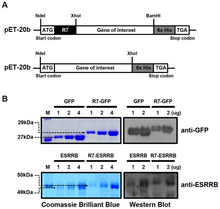
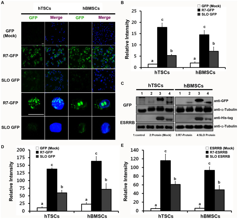
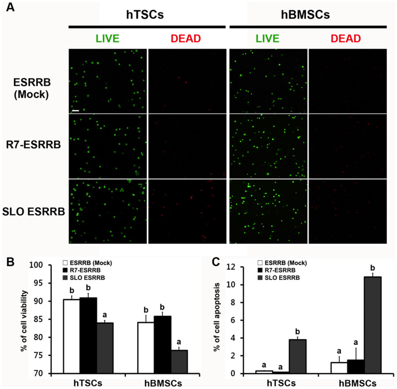
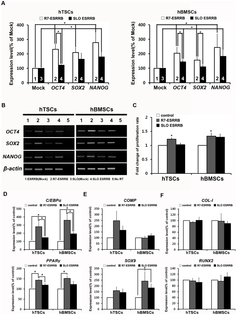
Delivery of proteins has been regarded as the safest and most useful application in therapeutic application of stem cells, because proteins can regulate gene expression transiently without any genomic alteration. However, it is difficult to accurately measure efficiency or quantity of intracellular protein uptake. Here, we performed a comparison study of cell-penetrating peptide (CPP)-conjugated protein delivery system using seven arginine and Streptolysin O (SLO)-mediated system. To compare CPP- and SLO-mediated protein delivery systems, we used GFP and ESRRB protein, which is known to regulate pluripotency-related genes, for delivery into human bone marrow stromal cells (hBMSCs) and human testicular stromal cells (hTSCs). We found that CPP-conjugated protein delivery was more efficient, lower cytotoxicity, and higher biological activity than SLO-mediated protein delivery system. These results suggest that delivery of CPP-conjugated proteins is an efficient tool for introducing biologically active proteins into cells and may have important implications in clinical cell-based therapy.


Figures

Figure 1
Figure 1. Characterization of recombinant proteins.
(A) Schematic diagrams of recombinant proteins with or without the CPP (R7)-conjugated vectors. (B) Identification of recombinant protein (GFP and ESRRB) and R7-conjugated protein (R7-GFP and R7-ESRRB) by Coomassie Brilliant Blue staining and Western blotting using specific antibodies against GFP and ESRRB. Full-length gel and blot images are available in Supplementary Figure 4. M, molecular weight marker.
Figure 2
Figure 2. Comparison of the efficiency of two different protein delivery systems (CPP- and SLO-mediated).
(A) Transduction of GFP and R7-GFP were detected by confocal microscopy. GFP or R7-GFP were visualized in green. Nuclei were counterstained with DAPI and the images were merged (The top 3 rows show 400× magnification and the bottom 2 rows show 1000× magnification plus 3× zoom). Scale bars represent 20 μm. (B) Relative intensity of GFP. (C) Quantification of delivery of protein (GFP and ESRRB) and CPP-conjugated protein (R7-GFP and R7-ESRRB) were confirmed by Western blot. Lane 1, non-treated control; Lane 2, mock protein control (GFP and ESRRB); Lane 3, CPP-conjugated protein (R7-GFP and R7-ESRRB); Lane 4, SLO-mediated protein (GFP and ESRRB). All samples were normalized to α-Tubulin. Full-length blot images are available in Supplementary Figure 4. Relative intensities are shown for both GFP (D) and ESRRB (E). Data are presented as means ± SEM of three replicates. a,b,c Different superscripts represent significant differences (p < 0.05).
Figure 3
Figure 3. Cytotoxicity assay.
(A) Cell viability assay; Live cells (green) and dead cells (red) were detected by fluorescence microscopy. (B) Live cells and dead cells were counted, and the ratio of live to dead cells was calculated. (C) Apoptosis was examined by the TUNEL assay. Scale bars represent 100 μm. Data are presented as means ± SEM of six (B) and three (C) replicates. a,b Different superscripts represent significant differences (p < 0.05).
Figure 4
Figure 4. ESRRB induced expression level of pluripotency-related genes, proliferation, and in vitro differentiation efficiency.
(A) Quantitative real-time RT-PCR and (B) RT-PCR analysis of pluripotency-related genes. Expression levels of OCT4, SOX2, and NANOG in hTSCs and hBMSCs were analyzed 24 hr after R7-ESRRB or SLO-mediated ESRRB delivery. Each gene was normalized to β-actin as a housekeeping control [1: ESRRB(Mock), 2: R7-ESRRB, 3: SLO(Mock), 4: SLO ESRRB]. (C) Altered proliferation rate of ESRRB delivered hTSCs and hBMSCs were calculated as fold change of population doubling number. In vitro differentiation analysis of ESRRB delivered cells was performed by quantitative real-time RT-PCR of (D) adipogenesis-specific markers C/EBPα and PPARγ, (E) chondrogenesis-specific markers COMP and SOX9, and (F) osteogenesis-specific markers COL-I and RUNX2. Data are presented as means ± SEM of four (A), six (B), three (C), and four (D–F) replicates. * p < 0.05.

References

    1. Liu Y. et al. Delivery of intact transcription factor by using self-assembled supramolecular nanoparticles. Angew Chem Int Ed Engl 50, 3058–62 (2011). - PMC - PubMed
    1. O'Malley J., Woltjen K. & Kaji K. New strategies to generate induced pluripotent stem cells. Curr Opin Biotechnol 20, 516–21 (2009). - PubMed
    1. Venkatesan J. K. et al. SOX9 gene transfer via safe, stable, replication-defective recombinant adeno-associated virus vectors as a novel, powerful tool to enhance the chondrogenic potential of human mesenchymal stem cells. Stem Cell Res Ther 3, 22 (2012). - PMC - PubMed
    1. Um S. H. Delivering Factors for Reprogramming a Somatic Cell to Pluripotency. Int J Stem Cells 5, 6–11 (2012). - PMC - PubMed
    1. Derossi D., Joliot A. H., Chassaing G. & Prochiantz A. The third helix of the Antennapedia homeodomain translocates through biological membranes. J Biol Chem 269, 10444–50 (1994). - PubMed

Publication types

MeSH terms