Review
. 2014 May:70:47-55.
doi: 10.1016/j.yjmcc.2014.02.017. Epub 2014 Mar 10.

Cardiac fibroblast in development and wound healing

Affiliations
Review

Cardiac fibroblast in development and wound healing

Arjun Deb et al. J Mol Cell Cardiol. 2014 May.

Abstract

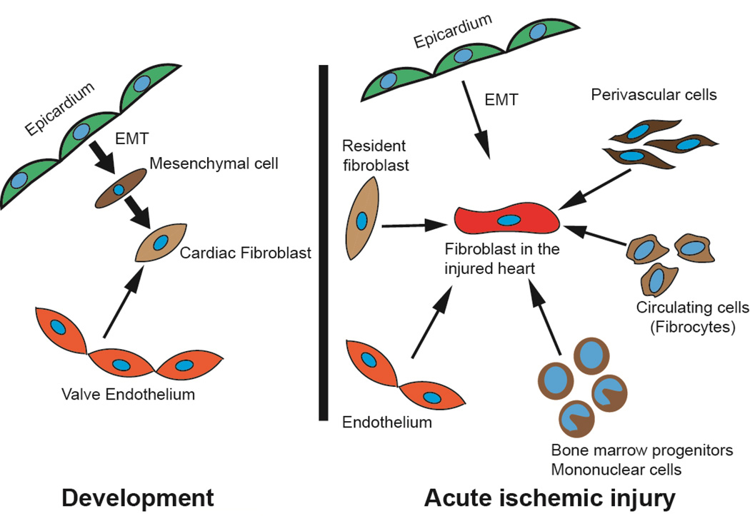
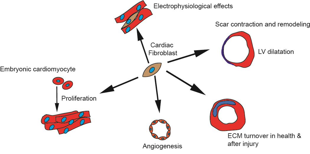
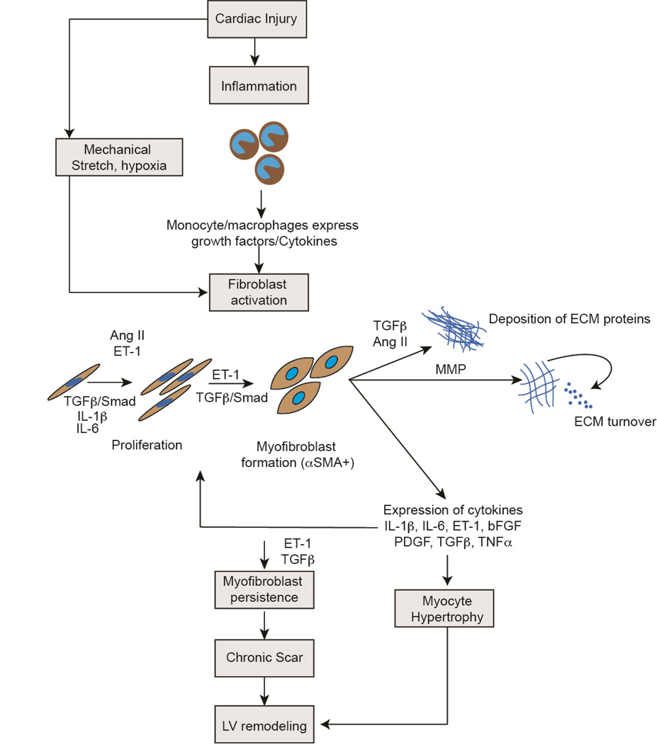
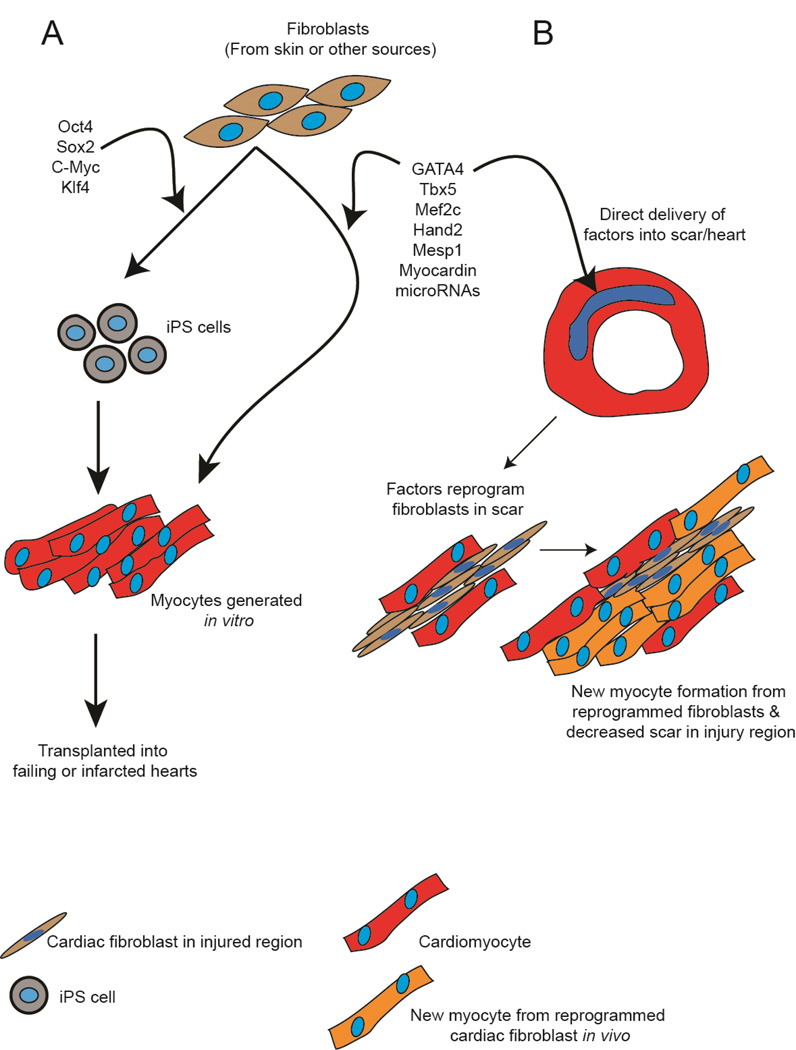
Cardiac fibroblasts are the most abundant cell type in the mammalian heart and comprise approximately two-thirds of the total number of cardiac cell types. During development, epicardial cells undergo epithelial-mesenchymal-transition to generate cardiac fibroblasts that subsequently migrate into the developing myocardium to become resident cardiac fibroblasts. Fibroblasts form a structural scaffold for the attachment of cardiac cell types during development, express growth factors and cytokines and regulate proliferation of embryonic cardiomyocytes. In post natal life, cardiac fibroblasts play a critical role in orchestrating an injury response. Fibroblast activation and proliferation early after cardiac injury are critical for maintaining cardiac integrity and function, while the persistence of fibroblasts long after injury leads to chronic scarring and adverse ventricular remodeling. In this review, we discuss the physiologic function of the fibroblast during cardiac development and wound healing, molecular mediators of activation that could be possible targets for drug development for fibrosis and finally the use of reprogramming technologies for reversing scar. This article is part of a Special Issue entitled "Myocyte-Fibroblast Signalling in Myocardium."

Keywords: Fibroblast; Fibrosis; Infarction; Remodeling; Repair; Wound healing.


Figures

Figure 1
Figure 1. Origins of cardiac fibroblasts during cardiac development and following acute ischemic injury
The cardiac fibroblast in the injured heart has diverse origins compared to the fibroblast in the developing heart.
Figure 2
Figure 2. Major functions of the fibroblast during cardiac development and wound healing
The fibroblast promotes proliferation of embryonic cardiomyocytes. It influences angiogenesis in the adult heart, regulates ECM turnover both in the adult uninjured heart and after acute injury. The fibroblast plays a pathophysiological role in scar contraction, adverse remodeling and ventricular dilatation and exerts electrophysiological effects.
Figure 3
Figure 3. Signaling pathways regulating pathophysiological effects of cardiac fibroblasts after cardiac injury
Following cardiac injury, monocyte/macrophage infiltrate at the site of injury release growth factors and cytokines that activate cardiac fibroblasts. Mechanical stretch and hypoxia in addition are potent activators of fibroblasts. TGFβ, ET-1, Ang II and other inflammatory cytokines such as IL-1β and IL-6 induce cardiac fibroblast proliferation, formation of myofibroblast, deposition of extracellular matrix and matrix turnover. The fibroblast itself expresses many of these pro-fibrotic cytokines that exert effects via autocrine/paracrine mechanisms. Cytokines expressed by cardiac fibroblasts also induce cardiomyocyte hypertrophy. Persistence of myofibroblasts in the injury region leads to continuing matrix turnover, formation of chronic scar and along with myocyte hypertrophy leads to adverse ventricular remodeling. (TGFβ: Transforming Growth Factor β, IL: Interleukin, ET-1: Endothelin 1, Ang II: Angiotensin II, bFGF: Basic Fibroblast Growth Factor, PDGF: Platelet Derived Growth Factor, TNFα: Tumor Necrosis Factor α, ECM: Extracellular matrix)
Figure 4
Figure 4. Targeting the fibroblast for cardiac regeneration
Fibroblasts can be reprogrammed into cardiomyocytes using cocktails of reprogramming factors. (A) In one approach, fibroblasts isolated from skin or other tissues of a ptient can be transduced with cocktails of transcription factors to yield induced pluripotent stem cells (iPS cells). Subsequently iPS cells generated from these fibroblasts can be differentiated in-vitro into cardiomyocytes that could be subsequently transplanted into failing or injured hearts. Alternatively combinations of factors can be used to directly reprogram the fibroblast to a cardiomyocyte in-vitro bypassing the iPS step and then generated cardiomyocytes would be transplanted in a similar manner. (B) In an alternative approach, combinations of factors can be directly delivered into the fibrotic region after cardiac injury. Cardiac fibroblasts in the injury region can directly reprogram into cardiomyocytes that potentially can integrate with existing cardiomyocytes to enhance cardiac function. By reprogramming fibroblasts in the injury region to form cardiomyocytes, this strategy would also decrease fibrosis and potentially ameliorate remodeling.

References

    1. Krenning G, Zeisberg EM, Kalluri R. The origin of fibroblasts and mechanism of cardiac fibrosis. J Cell Physiol. 2010;225:631–637. - PMC - PubMed
    1. Souders CA, Bowers SL, Baudino TA. Cardiac fibroblast: the renaissance cell. Circ Res. 2009;105:1164–1176. - PMC - PubMed
    1. Banerjee I, Fuseler JW, Price RL, Borg TK, Baudino TA. Determination of cell types and numbers during cardiac development in the neonatal and adult rat and mouse. Am J Physiol Heart Circ Physiol. 2007;293:H1883–H1891. - PubMed
    1. Zak R. Development and proliferative capacity of cardiac muscle cells. Circ Res. 1974;35(suppl II):17–26. - PubMed
    1. Zeisberg EM, Kalluri R. Origins of cardiac fibroblasts. Circ Res. 2010;107:1304–1312. - PMC - PubMed

MeSH terms

Substances