

Association of brain amyloid-β with cerebral perfusion and structure in Alzheimer's disease and mild cognitive impairment
- PMID: 24625697
- PMCID: PMC3999717
- DOI: 10.1093/brain/awu043
Association of brain amyloid-β with cerebral perfusion and structure in Alzheimer's disease and mild cognitive impairment
Abstract
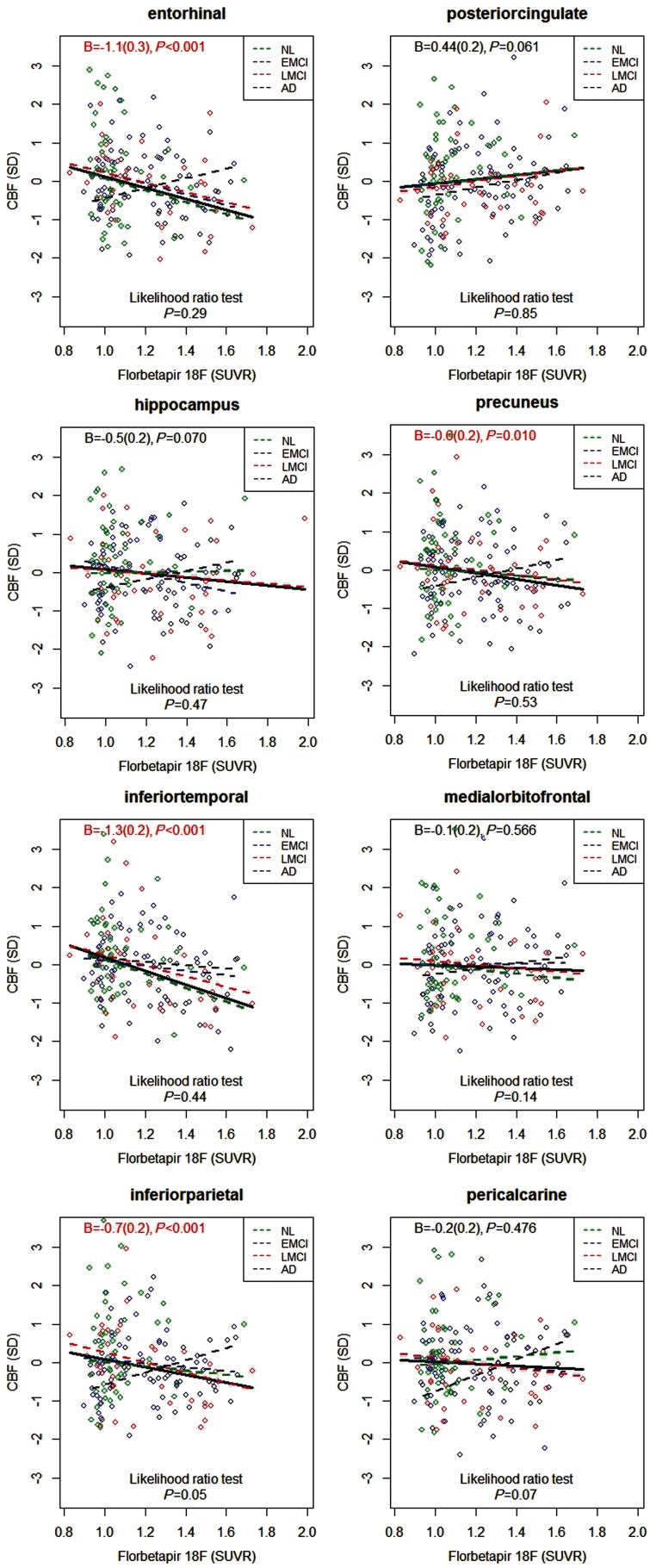
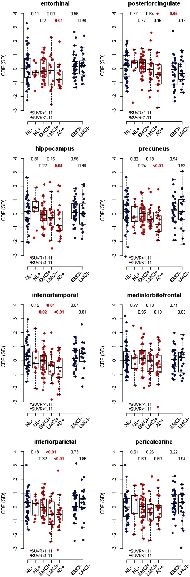
Patients with Alzheimer's disease have reduced cerebral blood flow measured by arterial spin labelling magnetic resonance imaging, but it is unclear how this is related to amyloid-β pathology. Using 182 subjects from the Alzheimer's Disease Neuroimaging Initiative we tested associations of amyloid-β with regional cerebral blood flow in healthy controls (n = 51), early (n = 66) and late (n = 41) mild cognitive impairment, and Alzheimer's disease with dementia (n = 24). Based on the theory that Alzheimer's disease starts with amyloid-β accumulation and progresses with symptoms and secondary pathologies in different trajectories, we tested if cerebral blood flow differed between amyloid-β-negative controls and -positive subjects in different diagnostic groups, and if amyloid-β had different associations with cerebral blood flow and grey matter volume. Global amyloid-β load was measured by florbetapir positron emission tomography, and regional blood flow and volume were measured in eight a priori defined regions of interest. Cerebral blood flow was reduced in patients with dementia in most brain regions. Higher amyloid-β load was related to lower cerebral blood flow in several regions, independent of diagnostic group. When comparing amyloid-β-positive subjects with -negative controls, we found reductions of cerebral blood flow in several diagnostic groups, including in precuneus, entorhinal cortex and hippocampus (dementia), inferior parietal cortex (late mild cognitive impairment and dementia), and inferior temporal cortex (early and late mild cognitive impairment and dementia). The associations of amyloid-β with cerebral blood flow and volume differed across the disease spectrum, with high amyloid-β being associated with greater cerebral blood flow reduction in controls and greater volume reduction in late mild cognitive impairment and dementia. In addition to disease stage, amyloid-β pathology affects cerebral blood flow across the span from controls to dementia patients. Amyloid-β pathology has different associations with cerebral blood flow and volume, and may cause more loss of blood flow in early stages, whereas volume loss dominates in late disease stages.
Keywords: Alzheimer’s disease; PET imaging; beta-amyloid; magnetic resonance imaging; perfusion imaging.
Figures
References
-
- Alexopoulos P, Sorg C, Förschler A, Grimmer T, Skokou M, Wohlschläger A, et al. Perfusion abnormalities in mild cognitive impairment and mild dementia in Alzheimer’s disease measured by pulsed arterial spin labeling MRI. Eur Arch Psychiatry Clin Neurosci. 2012;262:69–77. - PubMed
-
- Alsop DC, Detre JA, Grossman M. Assessment of cerebral blood flow in Alzheimer’s disease by spin-labeled magnetic resonance imaging. Ann Neurol. 2000;47:93–100. - PubMed
-
- Archer HA, Edison P, Brooks DJ, Barnes J, Frost C, Yeatman T, et al. Amyloid load and cerebral atrophy in Alzheimer’s disease: an 11C-PIB positron emission tomography study. Ann Neurol. 2006;60:145–7. - PubMed
Publication types
MeSH terms
Substances
Grants and funding
LinkOut - more resources
Full Text Sources
Other Literature Sources
Medical
