. 2014 Sep 15;210(6):954-63.
doi: 10.1093/infdis/jiu154. Epub 2014 Mar 12.

A role for the vacuolating cytotoxin, VacA, in colonization and Helicobacter pylori-induced metaplasia in the stomach

Affiliations

A role for the vacuolating cytotoxin, VacA, in colonization and Helicobacter pylori-induced metaplasia in the stomach

Jody A Winter et al. J Infect Dis. .

Abstract

Carriage of Helicobacter pylori strains producing more active (s1/i1) forms of VacA is strongly associated with gastric adenocarcinoma. To our knowledge, we are the first to determine effects of different polymorphic forms of VacA on inflammation and metaplasia in the mouse stomach. Bacteria producing the less active s2/i2 form of VacA colonized mice more efficiently than mutants null for VacA or producing more active forms of it, providing the first evidence of a positive role for the minimally active s2/i2 toxin. Strains producing more active toxin forms induced more severe and extensive metaplasia and inflammation in the mouse stomach than strains producing weakly active (s2/i2) toxin. We also examined the association in humans, controlling for cagPAI status. In human gastric biopsy specimens, the vacA i1 allele was strongly associated with precancerous intestinal metaplasia, with almost complete absence of intestinal metaplasia in subjects infected with i2-type strains, even in a vacA s1, cagA(+) background.

Keywords: Helicobacter pylori; SPEM; colonization; gastric cancer; pathogenesis; virulence.


Figures

Figure 1.
Figure 1.
Expression levels and activities of VacA from SS1 mutant series. A, Water extracts (7.5 µg total protein per lane) were Western blotted for VacA to confirm the presence or absence of toxin expression and relative quantities of toxin production. WT, wild type. B, RK13 cells were incubated for 4 hours in the presence of 75 µg/mL of the water extracts shown in A. The extent of vacuolation was quantified with light microscopy; vacuolating activity is presented as the mean ( ± standard deviation) proportion of vacuolated cells per field of view from ≥4 fields). C, Representative microscopic images of vacuolation.
Figure 2.
Figure 2.
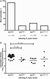
Expression of s2/i2 vacA promotes Helicobacter pylori colonization in mice. Mice were infected with SS1 expressing s1/i1, s1/i2, or s2/i2 or null for vacA. After 3 weeks, gastric colonization densities were determined by quantitative culture. Data shown for each group are combined from a minimum of 4 independent experiments. Data for vacA null group are combined from 2 independently produced vacA null mutants. A, Proportion of mice from which H. pylori was successfully recovered 3 weeks after inoculation. B, Colonization densities in the stomachs of infected mice. SS1s1i1 and SS1s1i2 colonized mice to 7.4-fold and 7.3-fold lower mean densities, respectively, than SS1s2i2. Of 26 mice inoculated with SS1null, bacteria were recovered from only 4 mice after 3 weeks, and the colonization densities were highly variable. Data were analyzed by means of 1-way analysis of variance with Tukey posttest. Horizontal lines indicate mean values for each experimental group. Abbreviation: CFU, colony-forming units.
Figure 3.
Figure 3.
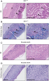
Infection with SS1s1i1 induces spasmolytic polypeptide-expressing metaplasia (SPEM) and inflammation in mouse gastric mucosa. A, Hematoxylin-eosin (HE)–stained gastric sections from a mouse 3 weeks after dosing with SS1s1i1 in Brucella broth showing inflammatory infiltrate (asterisk), loss of specialized gland cells, and marked expansion of cells with foamy cytoplasm indicative of mucinous metaplasia (arrows). B, Periodic acid–Schiff (PAS)/Alcian blue staining on gastric sections from SS1s1i1-dosed mice after 3 weeks of infection confirmed the replacement of specialized gland cells with mucin-containing cells (blue-staining acid mucin and purple-staining neutral mucin), consistent with SPEM [36]. C, Sections from a mouse dosed with Brucella broth only. D, SPEM was absent 3 weeks after dosing with Brucella broth. Panels A and B are representative of SPEM and inflammation scores of 3 (severe); panels C and D representative of inflammation scores of 0 (absent). Bar represents 200 µm for all panels.
Figure 4.
Figure 4.
Expression of s1/i1-type vacA induces more severe gastric inflammation and spasmolytic polypeptide-expressing metaplasia (SPEM) in the C57BL/6J mouse SS1 Helicobacter pylori infection model. Groups of 5–6 mice per study were inoculated with SS1 expressing s1/i1-, s1/i2-, or s2/i2-type VacA in Brucella broth. After 3 weeks, the severity of inflammation and SPEM in stomach sections were quantified histologically using an adapted Sydney scoring system. Inflammation and SPEM extent were quantified by measuring the length of inflamed or metaplastic corpus and expressing each as a proportion of the total corpus length examined. Data presented are combined from 2 independent studies; in each study, all 3 groups were examined in parallel. Horizontal lines indicate median values for each group. P values were calculated with 2-tailed Mann–Whitney tests.
Figure 5.
Figure 5.
Inoculation of C57BL/6J mice with SS1s1i1 or SS1s2i2 induces progressive inflammation and spasmolytic polypeptide-expressing metaplasia (SPEM). Groups of 5–10 mice were infected with SS1 expressing s2/i2 (A) or s1/i1 (B) type VacA in Brucella broth. After 1, 3, or 6 weeks, stomach sections were histologically scored using an adaptation of the Sydney system, and inflammation and SPEM extent expressed as percentage of total corpus length examined with scores of 1 (mild) to 3 (severe). The severity and extent of inflammation and SPEM increased progressively with infection time for both SS1s2i2 and SS1s1i1. Horizontal lines indicate median values for each group.
Figure 6.
Figure 6.
Carriage of vacA i1-type strains is associated with increased incidence of intestinal metaplasia in the human stomach. The severity of intestinal metaplasia in gastric biopsy specimens from patients infected with vacA s2i2- (n = 9), s1i2- (n = 23), or s1i1- (n = 53) type Helicobacter pylori strains was scored in a blinded fashion by a qualified histopathologist. Intestinal metaplasia was seen in a single patient infected with an i2-type strain (3%) but was present in 28% of patients infected only with i1-type strains (AC). The association between vacA i type and intestinal metaplasia remained when only patients infected with vacA s1-type strains (D) were included, and similar trends were observed in groups infected with vacA s1-type, cagA+ (E) and vacA s1-type, cagA strains (F). Data were analyzed using a Kruskal–Wallis test with Dunn posttest (A) and Fisher exact tests (BF).

References

    1. Kuipers EJ, Thijs JC, Festen HP. The prevalence of Helicobacter pylori in peptic ulcer disease. Aliment Pharmacol Ther. 1995;9(Suppl 2):59–69. - PubMed
    1. Ernst PB, Gold BD. The disease spectrum of Helicobacter pylori: the immunopathogenesis of gastroduodenal ulcer and gastric cancer. Ann Rev Microbiol. 2000;54:615–40. - PubMed
    1. El-Omar EM, Carrington M, Chow WH, et al. Interleukin-1 polymorphisms associated with increased risk of gastric cancer. Nature. 2000;404:398–402. - PubMed
    1. Kurata JH, Nogawa AN. Meta-analysis of risk factors for peptic ulcer. Nonsteroidal antiinflammatory drugs, Helicobacter pylori, and smoking. J Clin Gastroenterol. 1997;24:2–17. - PubMed
    1. Kusters JG, van Vliet AH, Kuipers EJ. Pathogenesis of Helicobacter pylori infection. Clin Microbiol Rev. 2006;19:449–90. - PMC - PubMed

Publication types