. 2014 Mar 13;9(3):e91169.
doi: 10.1371/journal.pone.0091169. eCollection 2014.

Surgical sutures filled with adipose-derived stem cells promote wound healing

Affiliations

Surgical sutures filled with adipose-derived stem cells promote wound healing

Ann Katharin Reckhenrich et al. PLoS One. .

Abstract

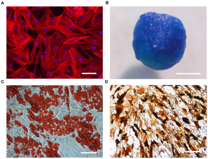
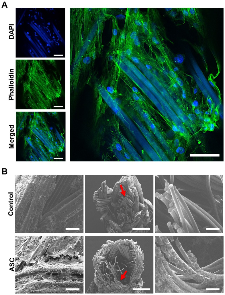
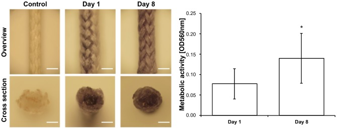
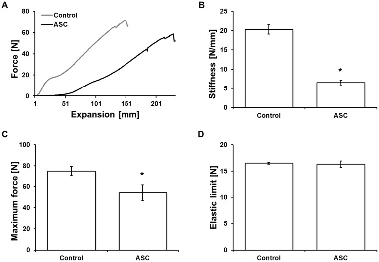
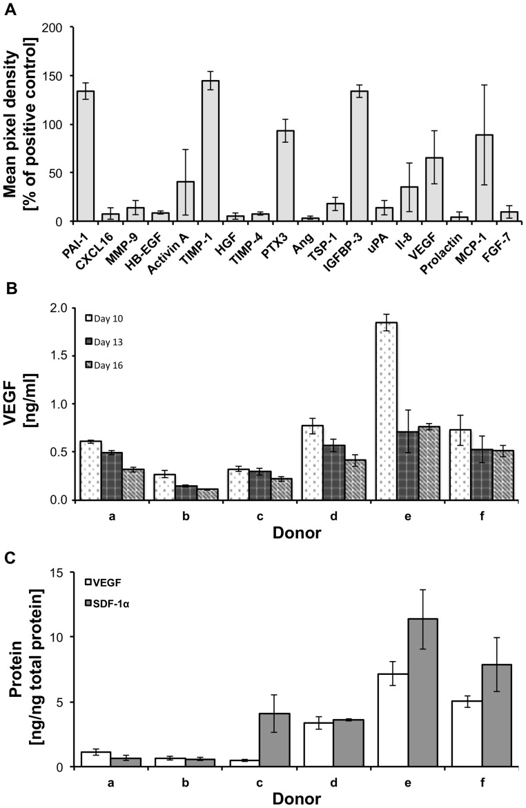
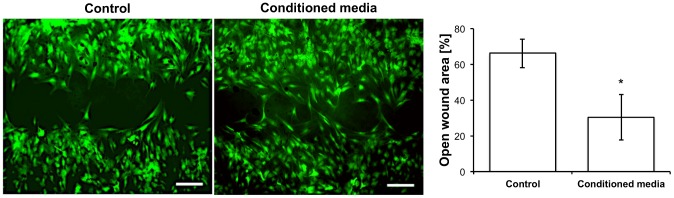
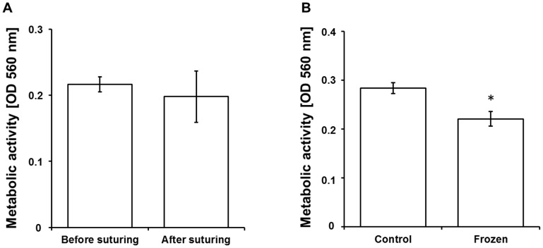
Delayed wound healing and scar formation are among the most frequent complications after surgical interventions. Although biodegradable surgical sutures present an excellent drug delivery opportunity, their primary function is tissue fixation. Mesenchymal stem cells (MSC) act as trophic mediators and are successful in activating biomaterials. Here biodegradable sutures were filled with adipose-derived mesenchymal stem cells (ASC) to provide a pro-regenerative environment at the injured site. Results showed that after filling, ASCs attach to the suture material, distribute equally throughout the filaments, and remain viable in the suture. Among a broad panel of cytokines, cell-filled sutures constantly release vascular endothelial growth factor to supernatants. Such conditioned media was evaluated in an in vitro wound healing assay and showed a significant decrease in the open wound area compared to controls. After suturing in an ex vivo wound model, cells remained in the suture and maintained their metabolic activity. Furthermore, cell-filled sutures can be cryopreserved without losing their viability. This study presents an innovative approach to equip surgical sutures with pro-regenerative features and allows the treatment and fixation of wounds in one step, therefore representing a promising tool to promote wound healing after injury.


Conflict of interest statement

Competing Interests: The authors have declared that no competing interests exist.

Figures

Figure 1
Figure 1. Cell characterization.
After isolation, cells adhered to tissue culture plastic showing a fibroblast-like morphology (A, DAPI (blue)/phalloidin (red) staining) and multipotency (B–D). Chondrogenic (B), adipogenic (C) and osteogenic (D) differentiation potential was confirmed by Alcian blue, Oil red O and von Kossa staining, respectively. Scale bars represent 100 μm in A, C, D and 1 mm in B.
Figure 2
Figure 2. Cellular distribution and attachment in the suture.
ASCs were filled into biodegradable sutures. Laser scanning microscopy (LSM, A) shows attached cells (DAPI, blue) along the suture surface interacting with each other (phalloidin, green) and the suture itself (DAPI, blue/phalloidin, green). The suture material is autofluorescent. As observed by scanning electron microscopy (SEM, B), cells were distributed throughout the surface (left) and the inner filaments (middle, right) of the suture. Scale bars represent 50 (A and B left), 500 (B, middle), and 100 μm (B, right).
Figure 3
Figure 3. Cell viability and proliferation in the suture.
The metabolic activity of ASCs in the suture was determined by MTT assay. Metabolically active cells (dark precipitate) were observed on the surface (overview) and in the inner cavity (cross section) of the suture at day 1 and 8 after seeding. Moreover, a significant increase of metabolic activity was observed from day 1 to 8. Scale bar represents 2 mm. p<0.05.
Figure 4
Figure 4. Biomechanical properties of ASC-filled sutures.
A force/expansion profile (A) was recorded to evaluate stiffness (B), maximum force (C), and the elastic limit (D) of ASC-filled sutures. Results show that stiffness and maximum force were significantly reduced when compared to cell-free sutures (control), but the elastic limit remained similar. p<0.001.
Figure 5
Figure 5. Cytokine release from ASC-filled sutures.
A cytokine array detected a multitude of different cytokines released from ASC-filled sutures (A). VEGF was constantly released to cell culture medium for at least 16 days (B). VEGF and SDF-1α were detected in protein extracts of ASC-filled sutures 16 days after seeding (C).
Figure 6
Figure 6. Biofunctionality of ASC-filled sutures.
Fresh media (control) and media conditioned with cell-filled sutures (conditioned) were tested in an in vitro wound healing assay (scratch assay) with ASCs. After 30 hours, a significant reduction in the open wound was observed when conditioned media was applied. Scale bars represent 500 μm. p<0.05.
Figure 7
Figure 7. Clinical perspective.
To determine the effect of mechanical stress on cell survival during suturing the metabolic activity of cell-filled sutures was determined before and after suturing in an ex vivo model using human skin, where no significant difference were observed (A). The potential to produce storable cell-filled sutures was evaluated. Therefore, the metabolic activity of ASCs in the suture was determined after freezing relative to unfrozen sutures. After freezing, 78% of the metabolic activity was preserved (B). p<0.001.

References

    1. Jones E, Yang X (2011) Mesenchymal stem cells and bone regeneration: current status. Injury 42: 562–568. - PubMed
    1. Sorrell JM, Caplan AI (2010) Topical delivery of mesenchymal stem cells and their function in wounds. Stem Cell Res Ther 1: 30. - PMC - PubMed
    1. Li H, Fu X (2012) Mechanisms of action of mesenchymal stem cells in cutaneous wound repair and regeneration. Cell Tissue Res 348: 371–377. - PubMed
    1. Brower J, Blumberg S, Carroll E, Pastar I, Brem H, et al. (2011) Mesenchymal stem cell therapy and delivery systems in nonhealing wounds. Adv Skin Wound Care 24: 524–532; quiz 533–524. - PubMed
    1. Choi KH, Choi BH, Park SR, Kim BJ, Min BH (2010) The chondrogenic differentiation of mesenchymal stem cells on an extracellular matrix scaffold derived from porcine chondrocytes. Biomaterials 31: 5355–5365. - PubMed

Publication types

MeSH terms

LinkOut - more resources