. 2014 Apr 4;344(6179):65-9.
doi: 10.1126/science.1250944. Epub 2014 Mar 13.

Polyhedra self-assembled from DNA tripods and characterized with 3D DNA-PAINT

Affiliations

Polyhedra self-assembled from DNA tripods and characterized with 3D DNA-PAINT

Ryosuke Iinuma et al. Science. .

Abstract

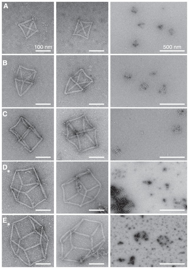
DNA self-assembly has produced diverse synthetic three-dimensional polyhedra. These structures typically have a molecular weight no greater than 5 megadaltons. We report a simple, general strategy for one-step self-assembly of wireframe DNA polyhedra that are more massive than most previous structures. A stiff three-arm-junction DNA origami tile motif with precisely controlled angles and arm lengths was used for hierarchical assembly of polyhedra. We experimentally constructed a tetrahedron (20 megadaltons), a triangular prism (30 megadaltons), a cube (40 megadaltons), a pentagonal prism (50 megadaltons), and a hexagonal prism (60 megadaltons) with edge widths of 100 nanometers. The structures were visualized by means of transmission electron microscopy and three-dimensional DNA-PAINT super-resolution fluorescent microscopy of single molecules in solution.


Figures

Fig. 1
Fig. 1. DNA-origami polyhedra
(A) Polyhedra self-assembled from DNA tripods with tunable inter-arm angles, and comparison of their sizes and molecular weights with selected previous polyhedra (structures 1 to 9) (details are provided in fig. S1). (B) Design diagram of a tripod. Cylinders represent DNA double helices. Details of the arm connection at the vertex are provided in fig. S2. (C) Cylinder model illustrating the connection between two tripod monomers. (D and E) Connection schemes for assembling (E) the tetrahedron and (D) other polyhedra (represented here by the cube design).
Fig. 2
Fig. 2. Self-assembly of DNA tripods and polyhedra
(A) Gel electrophoresis and (B) TEM images of the 60°-60°-60° (lane 1 in the gel) and 90°-90°-90° (lane 2) tripods. Gel lane 3 is a 1-kb ladder. Gel electrophoresis involves 1.5% native agarose gel and ice water bath. (C and D) Two schemes of connector designs and corresponding gel electrophoresis results. For each scheme, the strand model depicts the connection between two pairs of DNA duplexes. The number above a gel lane denotes the number of connected helices between two adjacent arms. Lane L is a 1-kb ladder, and lane S is a scaffold. Arrowheads indicate the bands corresponding to assembled cubes. (C) Scheme i: long (30 nt) connector (red) including a 2-nt sticky end. The complete 30-nt connector is only shown on the left, with a 28-nt segment anchored on the left helices and a 2-nt exposed sticky end available for hybridization with the 90°-90°-90° right neighbor (dashed circle depicts hybridization site). (D) Scheme ii: short (11 nt) connector including a 2-nt sticky end. (E) Assembly yields of the cubes, calculated as intensity ratio between a cube band and the corresponding scaffold band. (F) Agarose gel electrophoresis of the polyhedra. Lane 1 is the 90°-90°-90° monomer. Lanes 2 to 6 are polyhedra. Lane 7 is the assembly reaction containing tripods without struts. Lane 8 is the assembly reaction containing 90°-90°-90° tripods without vertex helices. Lane 9 is a 1-kb ladder. Gel bands corresponding to desired products are marked with arrowheads. Gel electrophoresis involves 0.8% native agarose gel and ice water bath.
Fig. 3
Fig. 3. TEM images of polyhedra
The zoomed-in (columns 1 and 2) and zoomed-out (column 3) images are shown for (A) the tetrahedron, (B) the triangular prism, (C) the cube, (D) the pentagonal prism, and (E) the hexagonal prism. Images of the tetrahedron, the triangular prism, and the cube were acquired from purified samples. Images of the pentagonal prism and the hexagonal prism were collected from crude samples (denoted with an asterisk). Scale bars are 100 nm in the zoomed-in TEM images and 500 nm in the zoomed-out images. Aggregates are clearly visible for unpurified samples [such as in (D), right].
Fig. 4
Fig. 4. 3D DNA-PAINT super-resolution fluorescence imaging of polyhedra
(A1) Staple strands at the vertices of each polyhedron were extended with single-stranded docking sequences for 3D DNA-PAINT super-resolution imaging. (A1 to E1) Schematics of polyhedra, with DNA-PAINT sites highlighted in green. (A2 to E2) 3D DNA-PAINT super-resolution reconstruction of typical polyhedra shown in the same perspective as depicted in A1 to E1. (A3 to E3) 2D x-y projection. (A4 to E4) 2D x-z projection. (A5 to E5) Height measurements of the polyhedra obtained from the cross-sectional histograms in the x-z projections. (F) A larger 2D super-resolution x-y projection view of tetrahedra and drift markers (bright individual dots). The diffraction-limited image is superimposed on the super-resolution image in the upper half. (G) Tilted 3D view of a larger-field-of-view image of the tetrahedron. Drift markers appear as bright individual dots. Scale bars, 200 nm. Color indicates height in the z direction.

References

    1. Chen JH, Seeman NC. Nature. 1991;350:631–633. - PubMed
    1. Winfree E, Liu F, Wenzler LA, Seeman NC. Nature. 1998;394:539–544. - PubMed
    1. Rothemund PW, Papadakis N, Winfree E. PLOS Biol. 2004;2:e424. - PMC - PubMed
    1. Rothemund PW. Nature. 2006;440:297–302. - PubMed
    1. Douglas SM, et al. Nature. 2009;459:414–418. - PMC - PubMed

Publication types