. 2014 Mar 13;5(3):e1118.
doi: 10.1038/cddis.2014.100.

Ubiquitin-like (UBX)-domain-containing protein, UBXN2A, promotes cell death by interfering with the p53-Mortalin interactions in colon cancer cells

Affiliations

Ubiquitin-like (UBX)-domain-containing protein, UBXN2A, promotes cell death by interfering with the p53-Mortalin interactions in colon cancer cells

S Sane et al. Cell Death Dis. .

Abstract

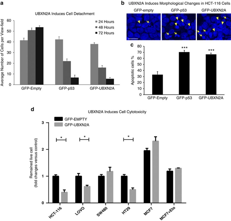
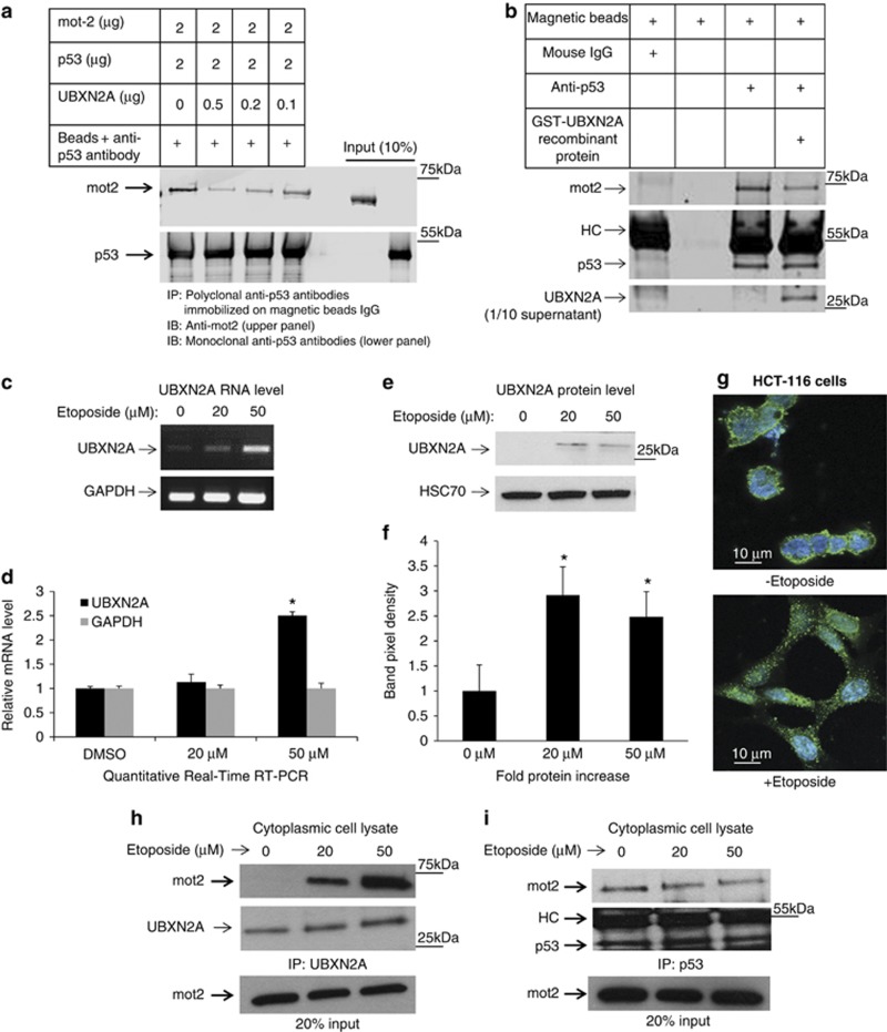
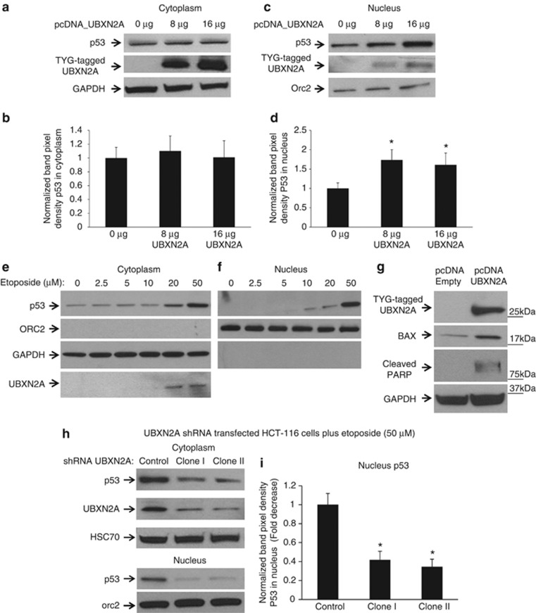
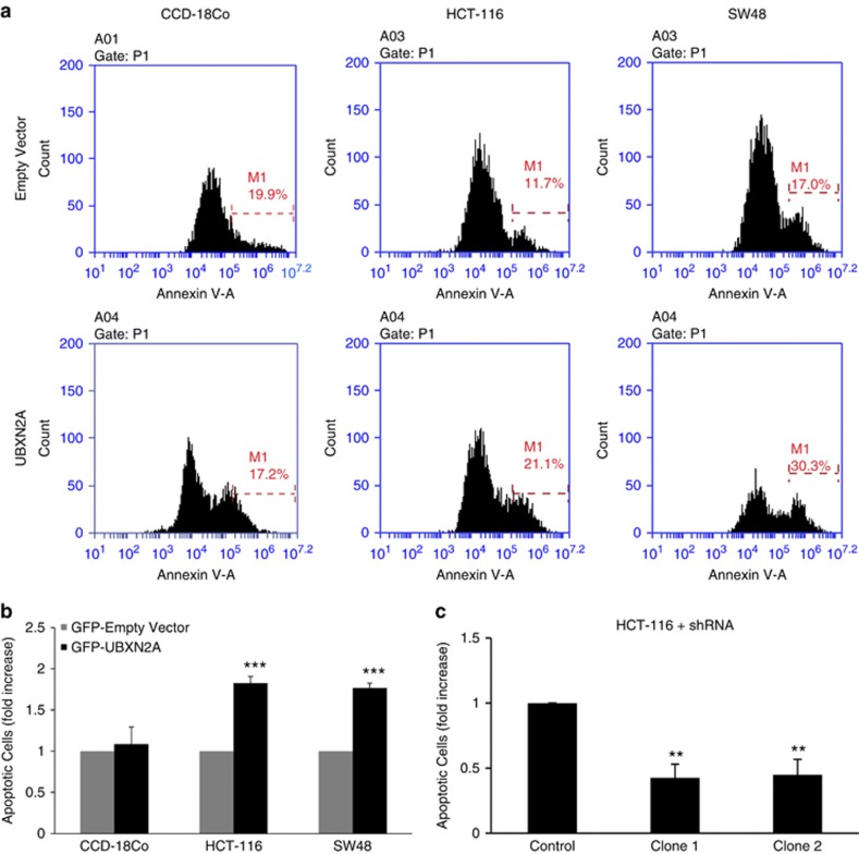
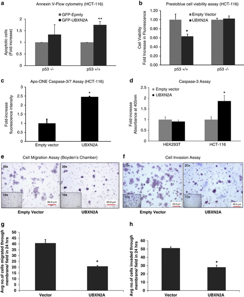
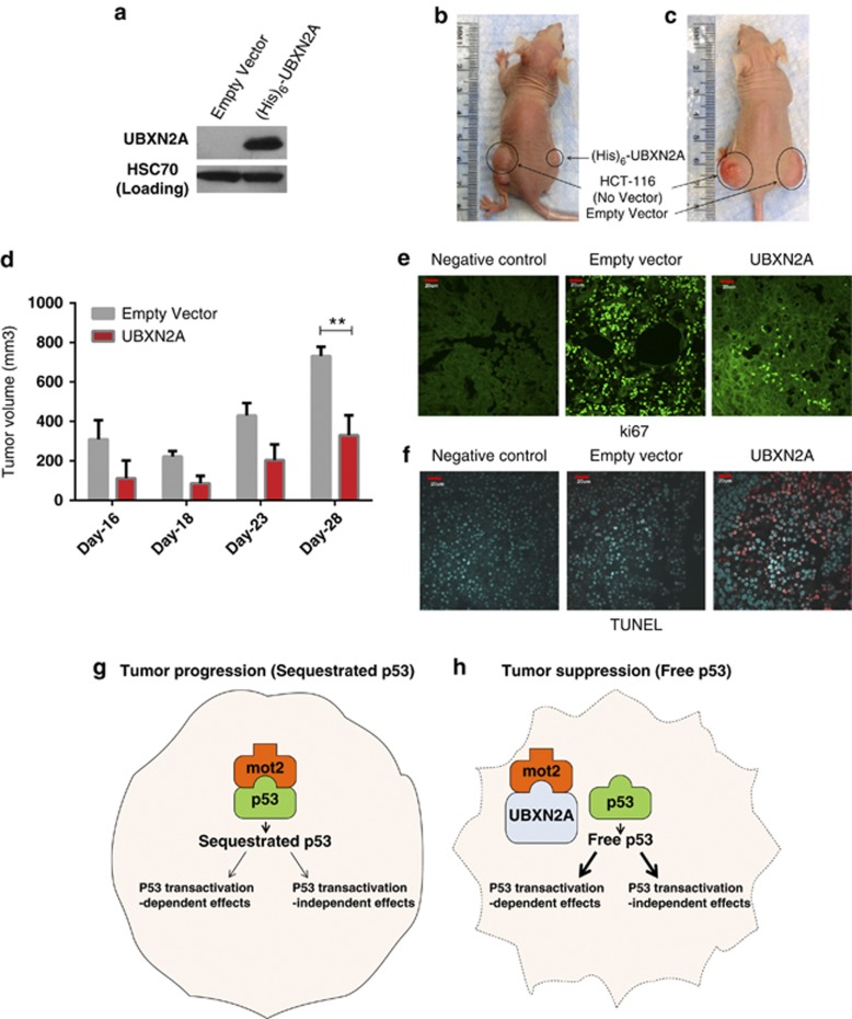
Mortalin (mot-2) induces inactivation of the tumor suppressor p53's transcriptional and apoptotic functions by cytoplasmic sequestration of p53 in select cancers. The mot-2-dependent cytoprotective function enables cancer cells to support malignant transformation. Abrogating the p53-mot-2 interaction can control or slow down the growth of cancer cells. In this study, we report the discovery of a ubiquitin-like (UBX)-domain-containing protein, UBXN2A, which binds to mot-2 and consequently inhibits the binding between mot-2 and p53. Genetic analysis showed that UBXN2A binds to mot-2's substrate binding domain, and it partly overlaps p53's binding site indicating UBXN2A and p53 likely bind to mot-2 competitively. By binding to mot-2, UBXN2A releases p53 from cytosolic sequestration, rescuing the tumor suppressor functions of p53. Biochemical analysis and functional assays showed that the overexpression of UBXN2A and the functional consequences of unsequestered p53 trigger p53-dependent apoptosis. Cells expressing shRNA against UBXN2A showed the opposite effect of that seen with UBXN2A overexpression. The expression of UBXN2A and its apoptotic effects were not observed in normal colonic epithelial cells and p53-/- colon cancer cells. Finally, significant reduction in tumor volume in a xenograft mouse model in response to UBXN2A expression was verified in vivo. Our results introduce UBXN2A as a home defense response protein, which can reconstitute inactive p53-dependent apoptotic pathways. Inhibition of mot-2-p53 interaction by UBXN2A is an attractive therapeutic strategy in mot-2-elevated tumors.


Figures

Figure 1
Figure 1
Cytotoxicity of UBXN2A in colon cancer cells with WT-p53. (a) HCT-116 cells were seeded at 30 000 cells per plate. Cells were transfected with GFP-empty vector, GFP-p53, and GFP-UBXN2A using the Neon transfection system (see Materials and Methods). Many GFP-UBXN2A cells were detached after the first 24 h. As GFP expression reached a maximum level after 24 h, cell detachment was studied after 24. 24, 48, and 72 h after transient transfection, detached cells were removed. Cell detachment was monitored using a Zeiss motorized inverted microscope and measuring the number of adherent live cells (green) using AxioVision software. The numbers of remaining cells were calculated in same five fields under the microscope for each experiment at 24, 48, and 72 h. The data from cell counting show a significant reduction of adherent cells in the presence of p53 or UBX2A in comparison with the empty vector after 72 h. These results suggest the UBXN2A-induced cell detachment due to apoptosis is comparable to that of p53. (b, c) In another set of experiments, HCT-116 cells transiently transfected with GFP-empty, GFP-p53, and GFP-UBXN2A were fixed and stained with DAPI. After 24 h, fluorescent microscopy observation demonstrates typical apoptotic morphology including condensation of the nuclear material (arrowheads) and formation of apoptotic bodies (arrows) when cells express p53 or UBXN2A (b). The bar chart represents the total number of the apoptotic cells counted in five fields for each groups after 24 h. Significant differences between different groups means and control value are indicated by ***P<0.001 (c). Scale bar=50 μm. (d) A cytotoxicity test with the crystal violet staining method was developed to determine cell viability in cancer cell lines transfected with GFP-UBXN2A. The Neon transfection system provides high efficiency of transfection (80–90%) and viability (90%) when electroporation parameters (voltage, pulse width, and number of pulses) are optimized for each individual cell line. Statistically significant cytotoxicity induced by UBXN2A was confirmed in HCT-116, LoVo, and HT-29 cell lines, while UBXN2A overexpression had no induction of cytotoxicity in SW480 cells (mutant p53) and MCF7 (caspase 3 deficient) breast cancer cells with and without suboptimal stress (Eto: etoposide 5 μM). Each value represents mean±S.E. of at least three independent experiments, and each experiment was performed in triplicate (*P<0.05)
Figure 2
Figure 2
UBXN2A interacts with mot-2. (a) HCT-116 cells were treated with etoposide (50 μM) for 24 h and cytoplasmic fractions were prepared. Samples were subjected to the iodixanol gradient centrifugation. The collected fractions (20 total) were analyzed by WB using the indicated antibodies. In HCT-116 cells, co-sedimentation of endogenous UBXN2A and mot-2 was predominantly observed in fractions 7-9 (black box). In contrast, P47 (UBXN2C) did not show co-sedimentation with mot-2, supporting a selective interaction between mot-2 and UBXN2A. In addition, UBXN2A showed co-sedimentation with its known partner, the protein p97 (CDC48). (b) HEK293T cells were transfected with increased amounts of pcDNA-(His)6-TYG-tagged UBXN2A. After 24 h, cell lysates were incubated with 50μl magnetic Dynabeads (His)6-Tag. UBXN2A pulls down endogenous mot-2 in a dose-dependent manner in HEK293T cells. (c, d) Using magnetic Dynabeads protein A, endogenous UBXN2A bound to immobilized anti-UBXN2A antibodies pulls down endogenous mot-2 in HCT-116 (knockout derivative (p53−/−) and wild-type (p53+/+)) colon cancer cells as well as mot-2 in human colon cancer tissues. We particularly observed pulled down mot-2 with tumors in d (Lane 4 versus 3). (e) Fractions shown in a were probed with anti-p53, HSP90α, and HSC70 antibodies. As expected, only some p53 proteins co-sediment with mot-2 (fractions 3–5). Instead, p53 showed strong co-sedimentation with fractions enriched in HSP90α, a known stabilizer of p53, in fractions 3–7. Fractions containing UBXN2A and mot-2 (a, fractions 7–9) have a low level of p53 (black box). As expected, another population of p53 proteins co-sedimented with HSC70, a known p53 regulator, in fractions 12–15. These results suggest that two distinct mot-2-containing complexes exist, one that sediments with p53 (fractions 3–5) and one that sediments with UBXN2A (fractions 7–9)
Figure 3
Figure 3
UBXN2A and p53 compete for binding to mot-2. (a) We investigated whether UBXN2A can decrease the association of p53 with mot-2 using an in vitro binding competition assay. First, recombinant human GST-p53 proteins bound to anti-p53 antibodies-IgG magnetic beads were incubated with human GST-mot-2 protein and increasing concentrations of human GST-UBXN2A recombinant proteins. Mot-2 proteins were eluted from the beads and analyzed by western blotting using an anti-mot-2 antibody. The same membrane was re-probed for p53 (lower panel) to show equivalent p53 in each IP. (b) The in vitro competition assay was further confirmed when the human GST-UBXN2A fusion proteins were incubated with cytosolic fractions enriched with mot-2 and p53 proteins (fractions 3-5, Figure 2e) of HCT-116 cells. The level of recombinant protein provided an ∼2.5:1 ratio of UBXN2A to endogenous mot-2. Cell lysates with and without UBXN2A were incubated with anti-p53 antibodies immobilized on magnetic Dynabeads protein G (Lanes 3 and 4). Beads with mouse IgG or beads alone were control groups in this experiment (lane 1 and 2). Western blotting showed that UBXN2A caused displacement of p53 binding from mot-2. These data support that UBXN2A and p53 compete for the mot-2. (c, d) A semiquantitative RT-PCR protocol (SuperScript III One-Step RT-PCR system) and quantitative real-time RT-PCR (d) showed etoposide enhances transcription of UBXN2A in HCT-116 cells. (e, f) At 24 h post-etoposide treatment (20 and 50 μM), total cell lysates were obtained, 50μg of protein was loaded in each lane, and the resulting blots were probed with an anti-UBXN2A antibody. UBXN2A's signal was normalized with the HSC70 signal (loading control). Together, these data confirmed UBXN2A expression is upregulated at the mRNA and protein levels upon genotoxic stress (P<0.05, n=3). DMSO was used as vehicle control. (g) Immunofluorescent staining reveals that UBXN2A shows a juxtanuclear staining characteristic of the ER/Golgi apparatus in the absence of etoposide. However, UBXN2A showed striking punctate cytoplasmic staining when HCT-116 cells incubated with etoposide (50 μM) for 24 h, suggesting a dynamic change in UBXN2A functional linkage networks in response to genotoxic stress. (h, i) The results presented in f indicate significant upregulation of UBXN2A in the cytoplasm with 20 and 50 μM etoposide after 24 h incubation. To verify whether this upregulated endogenous UBXN2A causes displacement of p53, we conducted a series of IP experiments. (h) Cytoplasmic lysates from e treated with no etoposide or 20 and 50 μM were subjected to IP using anti-UBXN2A (h) or anti-p53 (i) antibodies immobilized on magnetic IgG beads. Samples were resolved by 4–20% gradient SDS-PAGE and detected by WB using the indicated antibodies. The HC indicate the heavy immunoglobulin band. WB results indicate that UBXN2A binds to mot-2 in a etoposide dose-dependent manner, while the amount of mot-2 associated with p53 decreases simultaneously
Figure 4
Figure 4
UBXN2A induces p53 nuclear translocation in HCT-116 colon cancer cells. HCT-116 cells were transfected with the indicated amount of (His)6-TYG-tagged UBXN2A plasmid. Cytoplasmic (a) and nuclear (c) fractions were subjected to WB. The cell expression pattern of tagged UBXN2A proteins mimics the function of endogenous UBXN2A in the cytoplasm upon etoposide exposure illustrated in Figure 3. (b, d) UBXN2A is capable of significantly increasing the nuclear level of p53 in a dose-dependent manner (fold increase, n=11, *P<0.05). (e, f) HCT-116 cells were incubated with different concentrations of etoposide for 24 h. Cytoplasmic and nuclear protein lysates were prepared and subjected to western blot analysis to monitor p53 and UBXN2A protein levels. GAPDH and Orc-2 antibodies were used as cytoplasmic and nuclear markers, respectively. (g) HCT-116 cells were transfected with empty vector or (His)6-TYG-tagged UBXN2A plasmid. After 48 h, total cell lysates were prepared followed by WB using indicated antibodies. GAPDH was used as loading control. (h, i) Two lentiviral-based shRNAs were able to efficiently decrease the level of endogenous UBXN2A in HCT-116 cells treated for 24 h with 50 μM etoposide. WB of the nuclear cell lysates showed a significant decrease in nuclear p53 (i, n=8, *P<0.05). Taken together, these gain- and loss-of-function approaches in HCT-116 cells indicate that UBXN2A facilitates nuclear localization of transcriptionally active p53
Figure 5
Figure 5
UBXN2A overexpression induces apoptosis in colon cancer cell lines but not in normal, non-transformed cells. CCD-18Co, HCT-116, and SW48 were transfected with GFP-empty or GFP-UBXN2A. 48 h after transfection, cells were stained with Annexin V and early apoptosis was determined using flow cytometry as described in Materials and Methods. (a) Representative flow-cytometry analysis data from an Annexin V assay. The histograms show a comparison of the distribution of Annexin V positive cells (M1) after transient transfection of cells with GFP-empty vector or GFP-UBXN2A. The data were gated on GFP-UBXN2A positive cells prior to Annexin V analysis. (b) UBXN2A expression for 48 h significantly increased the number of apoptotic cells in HCT-116 and SW48 cell lines, while CCD-18Co normal colon cells were unaffected. (c) HCT-116 were transfected with scrambled shRNA or shRNA against UBXN2A (clone I and II). 48 h after silencing, superconfluent cultures of HCT-116 were analyzed by Annexin V assay using flow cytometry. Expression of GFP containing shRNA against UBXN2A resulted in 50% less apoptosis in comparison with cells transfected with scrambled shRNA. Values are expressed as mean (±S.E.M.) from three independent experiments (**P<0.01, ***P<0.001)
Figure 6
Figure 6
Induction of apoptosis by UBXN2A is p53 dependent and caspase-mediated in colon cancer cell lines. (a, b) HCT-116 (p53+/+) or HCT-116 (p53−/−) cells were transiently transfected with GFP-empty or GFP-UBXN2A for 48 h. An Annexin V apoptosis assay (a) and Prestoblue cell viability (b) assay show that overexpression of UBXN2A leads to a significant increase in cell apoptosis (c) and reduction of cell viability (d) in HCT-116 with WT-p53 (p53+/+). There was not a significant change between GFP-empty and GFP-UBXN2A in p53-KO cells (*P<0.05, **P<0.01). (c) HCT-116 cells were transfected with GFP-empty or GFP-UBXN2A. After 48 h, levels of caspase-3/7 activity (an indicator of apoptosis) in cells expressing GFP-empty or GFP-UBXN2A were measured using the Apo-ONE homogeneous caspase-3/7 assay kit. Results show 2.5-fold increase in caspase-3/7 activity in cells expressing GFP-UBXN2A relative to GFP-empty cells (*P<0.05). (d) Noncancerous HEK293T cells and HCT-116 colon cancer cell lines were transiently transfected with (His)6-TYG-empty or (His)6-TYG-UBXN2A vectors. A caspase-3 colorimetric assay revealed that UBXN2A exclusively activates caspase-3 only in cancer cells (*P<0.05). (e) Cell migration assay. HCT-116 empty-vector and HCT-116 UBXN2A-expressing cells (300 000 cells/well) were suspended in serum-free medium and seeded on cell culture inserts. After 24 h, cells that migrated through the membrane were stained and photographed at × 20 magnification. Cells were counted in five different fields and the average was plotted. (f) Invasion assay. HCT-116 empty vector and HCT-116 UBXN2A-expressing cells (300 000 cells/well) were suspended in serum-free medium and seeded on Matrigel coated inserts. After 24 h, cells that invaded through the Matrigel insert were stained and photographed at × 20 magnification. Cells were counted in five different fields, and the average was plotted. A representative micrograph (e, f) and quantification (g, h) of invaded cells are shown. The data show a significant decrease in migration/invasion of UBXN2A-expressing cells. For all the assays, data represent the mean of three experiments (Mean±S.E.M.) *P<0.05
Figure 7
Figure 7
Interaction of the SEP domain of UBXN2A with the p53-binding site (SBD domain) of mot-2 is sufficient to induce apoptosis. (a, b) Schematic diagram of UBXN2A and mot-2 protein domain structures. (cf) Comprehensive mapping of protein–protein interaction sites by Y2H method using α-galactosidase activity and nutritional selection verified that (i) WT-UBXN2A interacts with WT-mot-2, (ii) the SEP domain of UBXN2A is sufficient to interact with WT-mot-2, and (iii) a partial section of p53 binding site on the SBD domain of mot-2 (aa:438-506) is sufficient for binding to WT-UBXN2A. (g) An apoptosis assay using Annexin V staining followed by flow cytometry analysis confirmed that only the SEP domain of UBXN2A is required to induce apoptosis in HCT-116 cells, similar to full WT-UBXN2A. No increase in apoptosis was seen with the GFP-UBX domain. (h) HCT-116 cells were transfected with GFP-UBXN2A (WT) or its truncated forms (GFP-SEP or GFP-UBX domains). After 48 h, cells were subjected to a crystal violet cell cytotoxicity assay. Counting the remaining colonies showed both WT-UBXN2A and GFP-SEP domains significantly induce cell cytotoxicity (*P<0.05, n=3)
Figure 8
Figure 8
Overexpression of UBXN2A markedly reduces tumor growth in xenograft model. 7 × 10⁶ HCT-116 cells (p53+/+) expressing (His)6-TYG-UBXN2A or an empty vector were subcutaneously injected into nude mice in the lower flank (see Materials and Methods). (a) shows a portion of the transfected HCT-116 cells that was lysed and analyzed by western blotting for expression verification. (b, c) are representative of xenografts experiments with detectable expression of (His)6-tagged UBXN2A proteins. (d) Tumor growth was monitored on the indicated days. Results represent growth rate of tumor volume on the indicated days. Statistical comparisons were done by ANOVA followed by the Bonferroni post hoc test using the GraphPad Prism 6 (**P<0.01, n=3). Each data point is the mean tumor growth on the indicated day, and error bars show the standard error of mean. (e, f) Tumor tissue sections were subjected to immunohistochemical assay for Ki67 expression and TUNEL staining. Alexa-488 labeling of ki67 and 570 red fluorescent labeling for TUNEL assay were captured with the Olympus scanning confocal microscope using the Fluoview 1000 software. Negative controls were processed sections in the absence of primary antibody (e) or before proceeding to the TUNEL staining (f). Scale bar=20 μm. (g, h) The working model of p53 regulation by UBXN2A. Mot-2 protein has been shown to interact with p53 and inhibit its activation in cancerous cells. Upregulation of UBXN2A impedes mot-2-mediated inactivation of p53, which leads to the activation of p53

References

    1. Luo WI, Dizin E, Yoon T, Cowan JA. Kinetic and structural characterization of human mortalin. Protein Expr Purif. 2010;72:75–81. - PMC - PubMed
    1. Dundas SR, Lawrie LC, Rooney PH, Murray GI. Mortalin is over-expressed by colorectal adenocarcinomas and correlates with poor survival. J Pathol. 2005;205:74–81. - PubMed
    1. Takano S, Wadhwa R, Yoshii Y, Nose T, Kaul SC, Mitsui Y. Elevated levels of mortalin expression in human brain tumors. Exp Cell Res. 1997;237:38–45. - PubMed
    1. Wadhwa R, Takano S, Kaur K, Deocaris CC, Pereira-Smith OM, Reddel RR, et al. Upregulation of mortalin/mthsp70/Grp75 contributes to human carcinogenesis. Int J Cancer. 2006;118:2973–2980. - PubMed
    1. Ostermeyer AG, Runko E, Winkfield B, Ahn B, Moll UM. Cytoplasmically sequestered wild-type p53 protein in neuroblastoma is relocated to the nucleus by a C-terminal peptide. Proc Natl Acad Sci USA. 1996;93:15190–15194. - PMC - PubMed

Publication types

MeSH terms