. 2014 Mar 13;10(3):e1004006.
doi: 10.1371/journal.ppat.1004006. eCollection 2014 Mar.

Clonality of HTLV-2 in natural infection

Affiliations

Clonality of HTLV-2 in natural infection

Anat Melamed et al. PLoS Pathog. .

Abstract

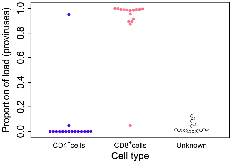
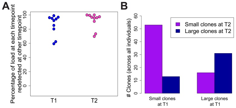
Human T-lymphotropic virus type 1 (HTLV-1) and type 2 (HTLV-2) both cause lifelong persistent infections, but differ in their clinical outcomes. HTLV-1 infection causes a chronic or acute T-lymphocytic malignancy in up to 5% of infected individuals whereas HTLV-2 has not been unequivocally linked to a T-cell malignancy. Virus-driven clonal proliferation of infected cells both in vitro and in vivo has been demonstrated in HTLV-1 infection. However, T-cell clonality in HTLV-2 infection has not been rigorously characterized. In this study we used a high-throughput approach in conjunction with flow cytometric sorting to identify and quantify HTLV-2-infected T-cell clones in 28 individuals with natural infection. We show that while genome-wide integration site preferences in vivo were similar to those found in HTLV-1 infection, expansion of HTLV-2-infected clones did not demonstrate the same significant association with the genomic environment of the integrated provirus. The proviral load in HTLV-2 is almost confined to CD8+ T-cells and is composed of a small number of often highly expanded clones. The HTLV-2 load correlated significantly with the degree of dispersion of the clone frequency distribution, which was highly stable over ∼8 years. These results suggest that there are significant differences in the selection forces that control the clonal expansion of virus-infected cells in HTLV-1 and HTLV-2 infection. In addition, our data demonstrate that strong virus-driven proliferation per se does not predispose to malignant transformation in oncoretroviral infections.


Conflict of interest statement

Niall Gormley is an employee of Illumina Inc, a public company that develops and markets systems for genetic analysis. This does not alter our adherence to all PLOS policies on sharing data and materials. The remaining authors have declared that no competing interests exist.

Figures

Figure 1
Figure 1. HTLV-2 infection is found almost exclusively in CD8+ T-cells.
Cryopreserved PBMCs from 28 HTLV-2-infected individuals were sorted by flow-cytometry into separate CD3+CD4+CD8 and CD3+CD4CD8+ cell populations. Integration site content was determined by high-throughput sequencing for both sorted populations and unsorted PBMCs. In the unsorted cells, integration sites were positively assigned to CD4+ or CD8+ cells based on the sorted fraction in which the same sites were found. The proportion of the load was calculated as the sum of the relative frequencies of the clones. Unknown – proportion of the load made up by clones that were not resampled in either the CD4+ or CD8+ fraction. Since the redetection of clones is most unlikely if proviral load is very low, only individuals in whom >100 proviruses were found are shown here.
Figure 2
Figure 2. HTLV-1 and HTLV-2 integrate in similar genomic environments.
Cells from 28 HTLV-2-infected and 16 HTLV-1-infected subjects were tested for genomic integration site preferences. (A) The ratio of the proportion of sites found in each chromosome (out of the total integration sites found for each virus) to the proportion of randomly generated (in silico) sites in the same chromosome. The yellow dashed line represents random sites (ratio = 1). (B) The number of histone marks (post-translational modifications) in three given windows across integration sites (for example, the 2k window incorporates 1,000 bases on either side of an integration site) compared to the number of histone marks in the same window across random sites. Statistical significance was assessed using the two-tailed Mann-Whitney test (* <0.05, ** <0.01, *** <0.001). (C) The odds ratio of integration within 1 kb of given ChIP-seq sites compared to random sites. The terms upstream and downstream here refer to the 5′ and 3′ sides of the integrated provirus, respectively. Statistical significance was assessed using Fisher's exact test (* <0.05, ** <0.01, *** <0.001).
Figure 3
Figure 3. HTLV-2 integration is highly oligoclonal, characterized by small numbers of expanded clones.
(A) Clonal distribution in representative subjects with HTLV-1 or HTLV-2 infection. The lowest observed, median and highest observed oligoclonality index values are shown. Each pie slice represents a single clone, proportional to relative abundance. Subjects with >100 proviruses identified are shown. OCI = oligoclonality index. (B) The observed number of clones in each subject with HTLV-1 or HTLV-2 infection (p<0.001, Mann-Whitney test). (C) The total number of clones in the blood was estimated using the DivE estimator (Laydon et al., manuscript submitted). Only samples containing sufficient information are shown. For each subject, the population size of infected cells in the blood was estimated based on the proviral load and average PBMC count. The estimated total number of clones in the blood was between 1 and 2 orders of magnitude lower in HTLV-2-infected subjects than in HTLV-1-infected subjects (p<0.001, Mann-Whitney test). (D) The oligoclonality index across all HTLV-1 -infected subjects compared to HTLV-2-infected subjects (p<0.001, Mann-Whitney test). (E) The percentage of the load maintained by clones observed only once compared between HTLV-1 and HTLV-2 (p<0.001, Mann-Whitney test).
Figure 4
Figure 4. HTLV-2 integration site and clonal expansion.
(A) The distribution of integration sites according to clonal abundance. Abundance was quantified by the number of copies estimated in a clone per 10,000 PBMCs (based on relative abundance and proviral load). Abundance bins are defined on a logarithmic scale. (B) The proportion of sites within 10 kb of a RefSeq gene for each abundance bin. A significant positive trend (p = 0.04, chi-squared test for trend) was detected for HTLV-1 but not for HTLV-2. (C) Oligoclonality index (OCI) versus log10(proviral load) for each virus. A strong positive correlation (p = 0.0015, Spearman's test) was detected between these parameters for HTLV-2 but not for HTLV-1(p = 0.681, Spearman's correlation). (D) The total number of unique integration sites (UIS) identified in each PBMC sample versus log10(proviral load) for each virus (p<0.001 for HTLV-1, p = 0.0019 for HTLV-2, Spearman's test).
Figure 5
Figure 5. HTLV-2 expanded clones are long-lived and stable.
Integration sites identified in PBMCs from an early time-point (T1, median = 9.9 years) were compared to those identified in the same HTLV-2-infected subjects at the present time-point (T2). (A) The percentage of proviral load (cumulative relative abundance) at each time-point represented by clones also present at the other time-point. (B) For clones found at both time-points, expanded clones (>1% of load) at any one time-point were significantly more likely to be expanded at both time-points (p<0.001, Fisher's exact test).

References

    1. Salemi M, Desmyter J, Vandamme AM (2000) Tempo and mode of human and simian T-lymphotropic virus (HTLV/STLV) evolution revealed by analyses of full-genome sequences. Mol Biol Evol 17: 374–386. - PubMed
    1. Akagi T, Shimotohno K (1993) Proliferative response of Tax1-transduced primary human T cells to anti-CD3 antibody stimulation by an interleukin-2-independent pathway. J Virol 67: 1211–1217. - PMC - PubMed
    1. Ross TM, Pettiford SM, Green PL (1996) The tax gene of human T-cell leukemia virus type 2 is essential for transformation of human T lymphocytes. J Virol 70: 5194–5202. - PMC - PubMed
    1. Roucoux DF, Murphy EL (2004) The epidemiology and disease outcomes of human T-lymphotropic virus type II. AIDS Rev 6: 144–154. - PubMed
    1. Gessain A, Cassar O (2012) Epidemiological Aspects and World Distribution of HTLV-1 Infection. Front Microbiol 3: 388. - PMC - PubMed

Publication types

MeSH terms