. 2014 Mar 13;10(3):e1003927.
doi: 10.1371/journal.ppat.1003927. eCollection 2014 Mar.

Inflammasome sensor NLRP1 controls rat macrophage susceptibility to Toxoplasma gondii

Affiliations

Inflammasome sensor NLRP1 controls rat macrophage susceptibility to Toxoplasma gondii

Kimberly M Cirelli et al. PLoS Pathog. .

Abstract

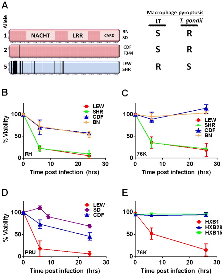
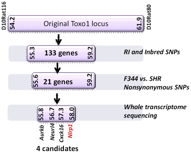
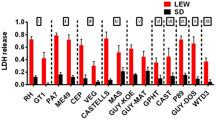
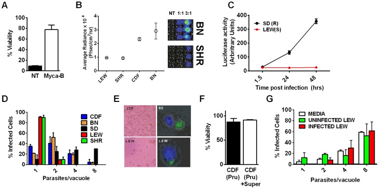
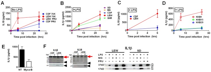
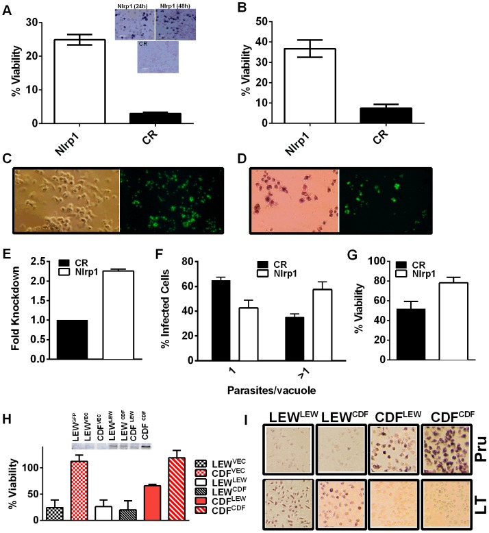
Toxoplasma gondii is an intracellular parasite that infects a wide range of warm-blooded species. Rats vary in their susceptibility to this parasite. The Toxo1 locus conferring Toxoplasma resistance in rats was previously mapped to a region of chromosome 10 containing Nlrp1. This gene encodes an inflammasome sensor controlling macrophage sensitivity to anthrax lethal toxin (LT) induced rapid cell death (pyroptosis). We show here that rat strain differences in Toxoplasma infected macrophage sensitivity to pyroptosis, IL-1β/IL-18 processing, and inhibition of parasite proliferation are perfectly correlated with NLRP1 sequence, while inversely correlated with sensitivity to anthrax LT-induced cell death. Using recombinant inbred rats, SNP analyses and whole transcriptome gene expression studies, we narrowed the candidate genes for control of Toxoplasma-mediated rat macrophage pyroptosis to four genes, one of which was Nlrp1. Knockdown of Nlrp1 in pyroptosis-sensitive macrophages resulted in higher parasite replication and protection from cell death. Reciprocally, overexpression of the NLRP1 variant from Toxoplasma-sensitive macrophages in pyroptosis-resistant cells led to sensitization of these resistant macrophages. Our findings reveal Toxoplasma as a novel activator of the NLRP1 inflammasome in rat macrophages.


Conflict of interest statement

The authors have declared that no competing interests exist.

Figures

Figure 1
Figure 1. NLRP1 sequence in inbred and RI rats correlates with rapid macrophage death.
(A) Sequence map of rat NLRP1 variants. This diagram was modified from . Vertical black lines indicate amino acid polymorphisms relative to the protein encoded by allele 1. Approximate NACHT, LRR and CARD domain locations relative to polymorphisms are shown. Macrophage sensitivity to LT-induced pyroptosis for the listed rat strains is from and Toxoplasma sensitivities are from this work. (B–E) Viability measurements for rat BMDMs from LEW, SHR (expressing NLRP1variant5); CDF, BN or SD (expressing NLRP1variant 1,2); or RI rat strains following infection with Toxoplasma Type I (RH) or Type II (76K or PRU) (MOI 3∶1) by MTT measurements. Data shown are average from three independent experiments with SD (triplicate wells/experiment/condition), except RI strains, which are averages from two experiments (triplicate wells/experiment/condition). Viability values were calculated relative to MTT measurements for uninfected control cells at each time point which were set at 100%. P-values comparing all NLRP1variant 1,2 -expressing strains to NLRP1variant 5 -expressing strains are <0.001.
Figure 2
Figure 2. Summary flow diagram for mapping of rat macrophage sensitivity to four candidate genes.
Methods for reducing the number of candidates at each stage are listed to the right and explained in detail in the Results section. Detailed SNPs and gene lists for each stage can be found in Supporting Figures S3 and S4 and Dataset S1.
Figure 3
Figure 3. NLRP1 variant-dependent rapid cell death is induced by many different parasite strains.
Viability as measured by LDH release for BMDMs from SD (NLRP11,2 variant) or LEW (NLRP15 variant) infected with strains representing global diversity for 24 h (Infection MOI 0.5–1 depending on strain, n = 4 wells/strain). P-values comparing LEW and SD <0.05 for all strains except MAS, CAST, GPHT and GUY-MAT.
Figure 4
Figure 4. NLRP1-variant dependent macrophage death depends on parasite invasion and controls parasite proliferation.
(A) Viability of LEW BMDMs infected with Mycalolide-treated (3 µM, 15 min) RH tachyzoites (MOI 1∶1) after 24 h as measured by MTS assay (P-value comparing Mycalolide group to untreated = 0.0002). (B, C) Radiance emission analyses of metabolically active, viable Type II Toxoplasma 76K parasites (B, graph MOI 3∶1, 6 h; inset shows representative plate from one experiment) or Type I RH parasites (C, MOI 1∶1 over 48 h) in BMDMs from different rat strains. P-value comparing NLRP1variant 1,2 expressing strains to NLRP1variant 5 expressing strains are <0.01 in I by t-test and <0.0001 in J by two-way ANOVA. (D) Number of parasites/vacuole in infected BMDMS (24 h, 3∶1 MOI) as assessed by microscopy is shown. CDF, BN infections were with 76K, and SD, LEW infections were with RH. Between 50–100 vacuoles counted per experiment. Average values from 3 experiments are shown for all strains, except SD (n = 2). P-values are <0.01 (two-way ANOVA) when comparing NLRP1variant 1, 2 expressing strains to NLRP1variant 5 expressing strains. (E) Left panels show light microscopy images of CDF and LEW monolayers infected with 76K (MOI 6∶1, 6 h). Right panels show fluorescence microscopy image of single SD and LEW BMDMs infected with RH (MOI 1∶1, 2 h). Blue is Hoechst stained nucleus, green are GFP-expressing parasites. Dividing parasites in SD cells (upper right) or a single parasite in LEW cells (lower right) are shown. (F) LEW BMDMs were infected with PRU (MOI 3;1) and at 5 h post infection culture supernatants from dying cells was spun, filtered and transferred to similarly infected (PRU, MOI 3∶1) CDF BMDMs. Viability of CDF BMDMs was assessed at 10 h post-infection by MTT staining. All values were calculated relative to uninfected control BMDMs (G) SD BMDMs were infected with RH parasites (2 h, MOI 1∶1), washed with PBS and medium replaced with fresh media, media from RH-infected (24 h, MOI 1∶1) or uninfected LEW BMDMs. Parasites/vacuole counted at 24 h. P-values >0.1 (ns) for comparison of any of three groups for 1, 2, 4 and 8 parasites/vacuole counts (by two-way ANOVA).
Figure 5
Figure 5. NLRP1-variant dependent cytokine cleavage and secretion.
IL-18 (A, C) and IL-1β (B, D) from LPS-primed (0.1 µg/ml, 1 h) (B, C, D) or unprimed (A) rat BMDMs following Toxoplasma infection (MOI 3∶1 for 76K and 3∶1 and 5∶1 for PRU). All infections are with strain 76K unless otherwise indicated with the additional exception that SD BMDMs in panel B were infected with PRU. Results shown are averages from three experiments with SD shown, except measurements for PRU infections in panel A which are the averages of four experiments, two with MOI 3∶1 and two with MOI 5∶1 and those for the RI rats, which are from two independent experiments (triplicate wells/experiment/time point). No IL-1 of IL-18 release was measurable from uninfected controls at any time point for any of the experiments in A–D. P-values in (A) comparing CDF and LEW groups in (A) and (C) are <0.001 by two-way ANOVA. In (B) and (D), all P-values comparing NLRP1 variant 5 expressing strains to the NLRP1 variant 1, 2-expressing strains are <0.001 in all comparison combinations, by two-way ANOVA (E) IL-1β measurements from LPS-primed LEW BMDMs infected with Mycalolide-treated (3 µM, 15 min) RH tachyzoites (MOI 1∶1) after 24 h; P-value comparing Mycalolide group to untreated is 0.0024 (F) Western blot analyses for IL-18 and IL-1β in cell lysates and culture supernatants (indicated by “S”) of 76K-infected CDF and LEW BMDMs (4 h, MOI 3∶1)(left panels) or PRU infected LEW and SD BMDM cell lysates (MOI 3∶1, 24 h)(right panels). NLRP3 agonist nigericin (40 µM, 4 h) was used as a positive control for inflammasome activation in the gel shown on the right. In the left pair of gels, supernatants (no concentration, mixed 1∶1 with SDS loading buffer) were loaded and Westerns were visualized using IR-dye conjugated secondary antibodies and the LiCOR Odyssey. Cell lysates were also run, with processed IL-1β and IL-18 shown with arrowheads in these gels, and pro-forms shown by red arrow. In the right gel, cell lysates are shown in Westerns visualized by chemiluminescence using a charge-coupled device camera. The unprocessed form of IL-1β is shown as the 37-kD band, and the mature form is labeled 17 kD.
Figure 6
Figure 6. Nlrp1 knockdown provides protection against Toxoplasma-induced pyroptosis and overexpression of NLRP1variant 5 sensitizes resistant macrophages.
(A) Viability of LEW BMDMs nucleofected with Nlrp1 siRNA pool or control siRNA (CR) 24 h or 48 h prior to infection with PRU (MOI 3∶1) as measured by MTT assay at 5 h post infection. Average from 6 separate nucleofection experiments (24 h n = 3, 48 h n = 3) are shown (triplicate wells/condition/experiment). P-values comparing Nlrp1 siRNA to controls is <0.001. Microscopy images of MTT stained nucleofected cells from representative 24 h and 48 h knockdown experiments are also shown. (B) Viability of LEW BMDMs nucleofected with Nlrp1 siRNA pool or control siRNA (CR) 36 h prior to infection with PRU (MOI 1∶1) as measured by MTT signal at 24 h post-infection. Average of 4 separate nucleofections are shown (triplicate wells/condition/nucleofection experiment) (C, D) Toxoplasma division in individually surviving nucleofected LEW BMDMs from (B) at 24 h post-infection. In C cells were fixed prior to microscopy, while in D cells were MTT-stained and fluorescence microscopy performed with no fixing. Note that all non-transfected or control siRNA transfected LEW macrophages which have succumbed are not present in these fields (detached by 24 h), while the MTT-negative ghosts and organelles of these lysed cells can be seen in parallel experiments at the earlier 5–6 h time, as shown in panel A. (E–G) Knockdown by the alternative lentiviral shRNA method was confirmed in LEW BMDMs by qPCR (E) and parasites per vacuole counts (F) and viability by MTS assay (G) were assessed in Nlrp1-knockdown LEW BMDMs after RH strain infection (MOI 0.5∶1). P-values by t-test comparing knockdown to controls is 0.03 for C and 0.01 for D. (H) Viability of LEW and CDF BMDMs nucleofected with full length HA-tagged NLRP1 constructs at −24 h prior to infection with PRU (MOI 5∶1) was measured by MTT assay at 5 h post-infection. Cell lysates from nucleofected cells were made at 32 h post-transfection and analyzed by Western using anti-HA antibody. Superscripts indicate the NLRP1 construct or vector that was transfected into the cell. Graph shows average from two nucleofection studies, with duplicate wells/condition/experiment. Lysates are from one of these nucleofections. There is no significant difference between any of the nucleofected LEW cells. The P-value comparing the CDF cells (expressing NLRP1variant 2) transfected with LEW (NLRP1variant 5) to CDF cells nucleofected with vector or CDF (NLRP1variant 2) is <0.0005. Presence of MTT-negative cells was also verified by microscopy for each well. Similar data is also shown in Figure S8, with anthrax LT control treatments. (I) Representative microscopy images of MTT viability staining for LEW and CDF BMDMs nucleofected with full length HA-tagged NLRP1 constructs −36 h prior to infection with PRU (MOI 3∶1) or treatment with LT (PA + LF, each at 1 µg/ml). MTT staining was performed on Toxoplasma-infected cells at 8 h post-infection and on LT-treated cells at 5 h post-infection. Superscripts indicate the NLRP1 construct or vector that was transfected into the cell.

References

    1. Melo MB, Jensen KD, Saeij JP (2011) Toxoplasma gondii effectors are master regulators of the inflammatory response. Trends Parasitol 27: 487–495. - PMC - PubMed
    1. Cavailles P, Sergent V, Bisanz C, Papapietro O, Colacios C, et al. (2006) The rat Toxo1 locus directs toxoplasmosis outcome and controls parasite proliferation and spreading by macrophage-dependent mechanisms. Proc Natl Acad Sci U S A 103: 744–749. - PMC - PubMed
    1. Sergent V, Cautain B, Khalife J, Deslee D, Bastien P, et al. (2005) Innate refractoriness of the Lewis rat to toxoplasmosis is a dominant trait that is intrinsic to bone marrow-derived cells. Infect Immun 73: 6990–6997. - PMC - PubMed
    1. Newman ZL, Printz MP, Liu S, Crown D, Breen L, et al. (2010) Susceptibility to anthrax lethal toxin-induced rat death is controlled by a single chromosome 10 locus that includes rNlrp1. PLoS Pathog 6: e1000906. - PMC - PubMed
    1. Pravenec M, Gauguier D, Schott JJ, Buard J, Kren V, et al. (1996) A genetic linkage map of the rat derived from recombinant inbred strains. Mamm Genome 7: 117–127. - PubMed

Publication types