

TMJ response to mandibular advancement surgery: an overview of risk factors
- PMID: 24626243
- PMCID: PMC3908759
- DOI: 10.1590/1678-775720130056
TMJ response to mandibular advancement surgery: an overview of risk factors
Abstract
Objective: In order to understand the conflicting information on temporomandibular joint (TMJ) pathophysiologic responses after mandibular advancement surgery, an overview of the literature was proposed with a focus on certain risk factors.
Methods: A literature search was carried out in the Cochrane, PubMed, Scopus and Web of Science databases in the period from January 1980 through March 2013. Various combinations of keywords related to TMJ changes [disc displacement, arthralgia, condylar resorption (CR)] and aspects of surgical intervention (fixation technique, amount of advancement) were used. A hand search of these papers was also carried out to identify additional articles.
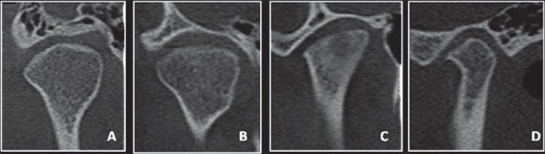
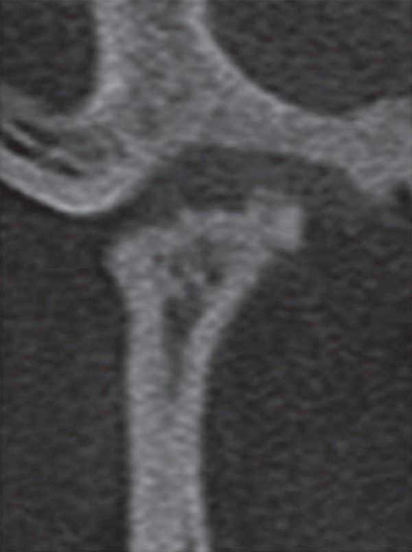
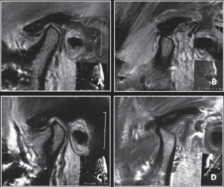
Results: A total of 148 articles were considered for this overview and, although methodological troubles were common, this review identified relevant findings which the practitioner can take into consideration during treatment planning: 1- Surgery was unable to influence TMJ with preexisting displaced disc and crepitus; 2- Clicking and arthralgia were not predictable after surgery, although there was greater likelihood of improvement rather than deterioration; 3- The amount of mandibular advancement and counterclockwise rotation, and the rigidity of the fixation technique seemed to influence TMJ position and health; 4- The risk of CR increased, especially in identified high-risk cases.
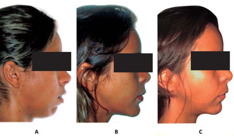
Conclusions: Young adult females with mandibular retrognathism and increased mandibular plane angle are susceptible to painful TMJ, and are subject to less improvement after surgery and prone to CR. Furthermore, thorough evidenced-based studies are required to understand the response of the TMJ after mandibular advancement surgery.
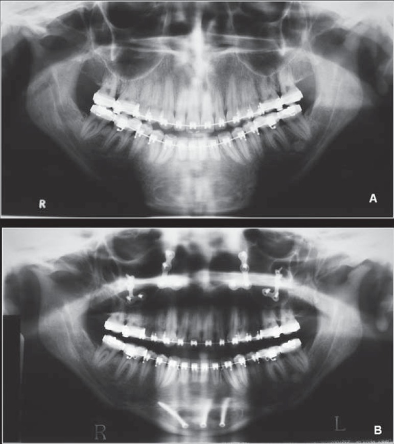
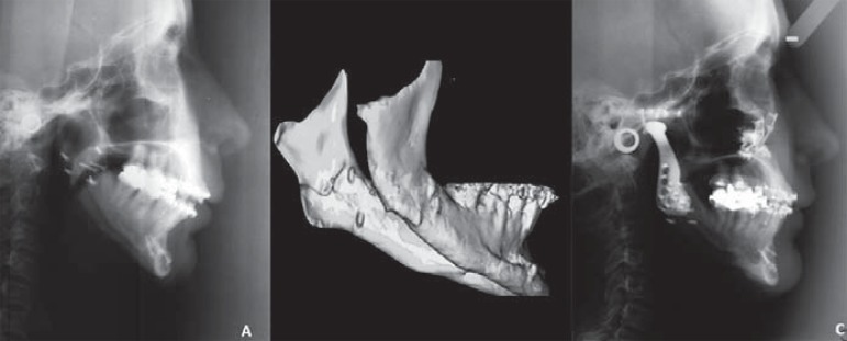
Figures
References
-
- Abrahamsson C, Ekberg E, Henrikson T, Bondemark L. Alterations of temporomandibular disorders before and after orthognathic surgery: a systematic review. Angle Orthod. 2007;77:729–734. - PubMed
-
- Abrahamsson C, Ekberg E, Henrikson T, Nilner M, Sunzel B, Bondemark L. TMD in consecutive patients referred for orthognathic surgery. Angle Orthod. 2009;79:621–627. - PubMed
-
- Ahmad M, Hollender L, Anderson Q, Kartha K, Ohrbach R, Truelove EL, et al. Research diagnostic criteria for temporomandibular disorders (RDC/TMD): development of image analysis criteria and examiner reliability for image analysis. Oral Surg Oral Med Oral Pathol Ora Radiol Endod. 2009;107:844–860. - PMC - PubMed
-
- Alder ME, Deahl ST, Matteson SR, Van Sickels JE, Tiner BD, Rugh JD. Short-term changes of condylar position after sagittal split osteotomy for mandibular advancement. Oral Surg Oral Med Oral Pathol Oral Radiol Endod. 1999;87:159–165. - PubMed
-
- Alexander G, Stivers M. Control of the proximal segment during application of rigid internal fixation of sagittal split osteotomy of the mandible. J Oral Maxillofac Surg. 2003;61:1113–1114. - PubMed
Publication types
MeSH terms
Grants and funding
LinkOut - more resources
Full Text Sources
Other Literature Sources
Medical
