

Editorial
doi: 10.1590/S1806-37132014000100001.
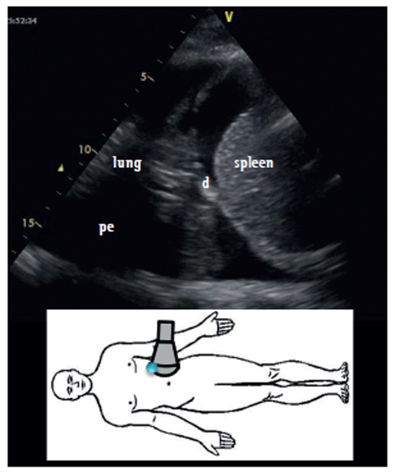
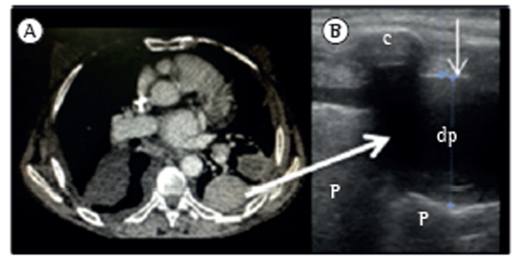
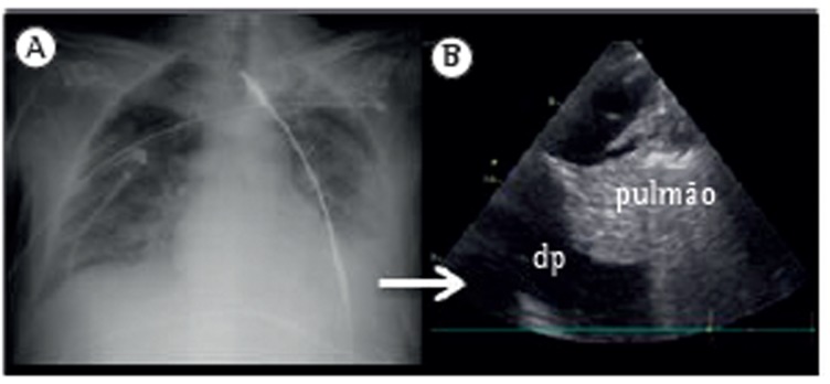
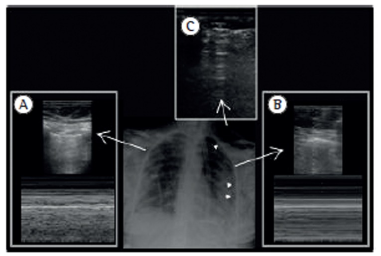
Lung ultrasound in the evaluation of pleural effusion
[Article in
English,
Portuguese]
Affiliations
- PMID: 24626263
- PMCID: PMC4075927
- DOI: 10.1590/S1806-37132014000100001
Item in Clipboard
Editorial
Lung ultrasound in the evaluation of pleural effusion
[Article in
English,
Portuguese]
J Bras Pneumol.
2014 Jan-Feb.
No abstract available
Figures


Comment on
-
Can ultrasound guidance reduce the risk of pneumothorax following thoracentesis?J Bras Pneumol. 2014 Jan-Feb;40(1):6-12. doi: 10.1590/S1806-37132014000100002. J Bras Pneumol. 2014. PMID: 24626264 Free PMC article. Clinical Trial.
References
-
- Koenig SJ, Narasimhan M, Mayo PH. Thoracic ultrasonography for the pulmonary specialist. Chest. 2011;140(5):1332–1341. http://dx.doi.org/10.1378/chest.11-0348 - DOI - PubMed
-
- Yu CJ, Yang PC, Wu HD, Chang DB, Kuo SH, Luh KT. Ultrasound study in unilateral hemithorax opacification. Image comparison with computed tomography. Am Rev Respir Dis. 1993;147(2):430–434. http://dx.doi.org/10.1164/ajrccm/147.2.430 - DOI - PubMed
-
- Lichtenstein D, Goldstein I, Mourgeon E, Cluzel P, Grenier P, Rouby JJ. Comparative diagnostic performances of auscultation, chest radiography, and lung ultrasonography in acute respiratory distress syndrome. Anesthesiology. 2004;100(1):9–15. http://dx.doi.org/10.1097/00000542-200401000-00006 - DOI - PubMed
-
- Blackmore CC, Black WC, Dallas RV, Crow HC. Pleural fluid volume estimation: a chest radiograph prediction rule. Acad Radiol. 1996;3(2):103–109. http://dx.doi.org/10.1016/S1076-6332(05)80373-3 - DOI - PubMed
Publication types
MeSH terms
LinkOut - more resources
Full Text Sources
Other Literature Sources
Medical
