. 2014 Aug 1;23(15):4015-23.
doi: 10.1093/hmg/ddu115. Epub 2014 Mar 13.

Disruption of the methyltransferase-like 23 gene METTL23 causes mild autosomal recessive intellectual disability

Affiliations

Disruption of the methyltransferase-like 23 gene METTL23 causes mild autosomal recessive intellectual disability

Marie Bernkopf et al. Hum Mol Genet. .

Abstract

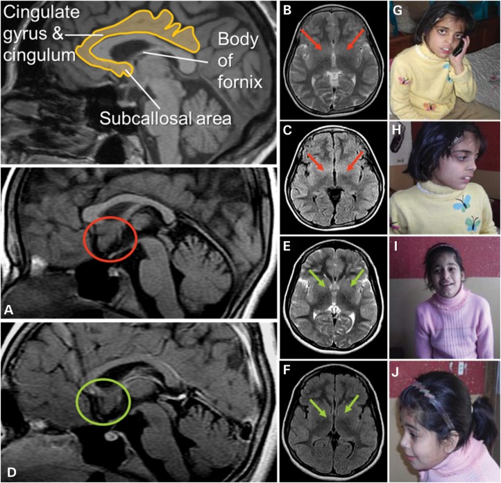
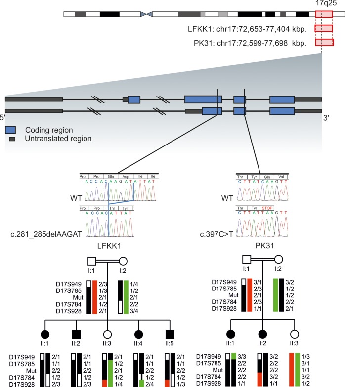
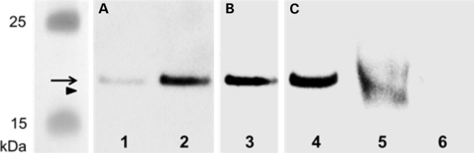
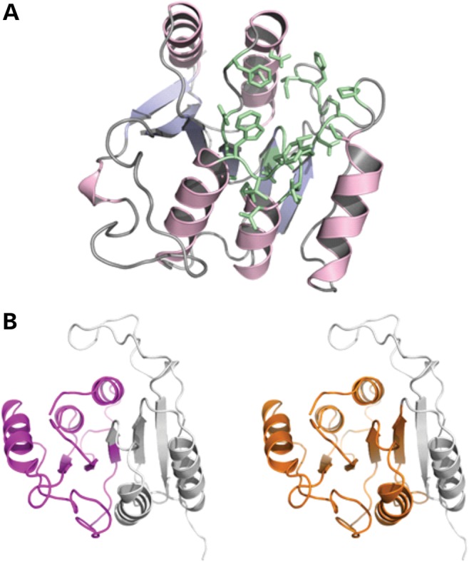
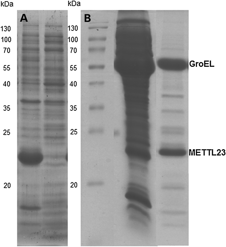
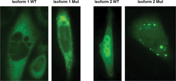
We describe the characterization of a gene for mild nonsyndromic autosomal recessive intellectual disability (ID) in two unrelated families, one from Austria, the other from Pakistan. Genome-wide single nucleotide polymorphism microarray analysis enabled us to define a region of homozygosity by descent on chromosome 17q25. Whole-exome sequencing and analysis of this region in an affected individual from the Austrian family identified a 5 bp frameshifting deletion in the METTL23 gene. By means of Sanger sequencing of METTL23, a nonsense mutation was detected in a consanguineous ID family from Pakistan for which homozygosity-by-descent mapping had identified a region on 17q25. Both changes lead to truncation of the putative METTL23 protein, which disrupts the predicted catalytic domain and alters the cellular localization. 3D-modelling of the protein indicates that METTL23 is strongly predicted to function as an S-adenosyl-methionine (SAM)-dependent methyltransferase. Expression analysis of METTL23 indicated a strong association with heat shock proteins, which suggests that these may act as a putative substrate for methylation by METTL23. A number of methyltransferases have been described recently in association with ID. Disruption of METTL23 presented here supports the importance of methylation processes for intact neuronal function and brain development.


Figures

Figure 1.
Figure 1.
Magnetic resonance images: Two independent experts in neuroimaging (Christian Enzinger, Franz Fazekas) inspected the MRI scans blinded to clinical information and detected increased volume of the subcallosal area (SA), which was particularly evident in patient PK31 II:2 (A: red circles; in comparison with patient II:1 in D: green circles). On axial T2-weighted and FLAIR sequences, this led to decreased identifiability of the basal ganglia (B, C red arrows (II:2); compared with E and F (II:1), green arrows). The SA is commonly attributed to the limbic system, belongs to a phylogenetic old-brain area, is located in the vicinity of strategically important areas such as the hypothalamus and mamillar bodies and has been implicated in affect regulation. Interestingly, clinical information revealed that patient II:2 had demonstrated aggressive behavior and disturbed impulse control. Photos of PK31 II:2 (G and H) and II:1 (I and J) are shown.
Figure 2.
Figure 2.
Ideogram of chromosome 17 is shown. Localization, indicated by red rectangles and base pair positions (referring to hg19) defining the homozygous regions of family FLKK1 and PK31 are given. The exon/intron structure of METTL23 is shown: blue blocks represent coding regions and gray blocks represent untranslated regions, introns are shown as dark lines. Black vertical lines indicate the position of the mutations for LFKK1 and PK31. Electropherograms show the homozygous familial mutations in METTL23 for LFKK1 and PK31 compared with WT sequences. Haplotypes and simplified pedigrees of LFKK1 and PK31 are shown. The disease haplotype is indicated by black bars. All alleles are recoded. Mutation status (mut) of all tested individuals is indicated by 1 for WT and 2 for mutated.
Figure 3.
Figure 3.
Western blot for METTL23 protein: Purified METTL23-His8-tag protein (expressed in E. coli) was loaded in lane 1 (50 ng) and 2–4 (100 ng). METTL23 proteins were immunoprecipitated from lymphoblastoid cells from healthy control (lane 5) and METTL23-mutant patient PK31 II:1 (lane 6) using polyclonal anti-METTL23. Immunoreactive bands were visualized using (A) rabbit polyclonal anti-METTL23-His8-tag antiserum, (B) rabbit polyclonal anti-His-tag (C-terminal) antibody and (C) sequence-specific anti-METTL23 (C17orf95) antibody as primary antibodies. Arrow (METTL23-His8-tag protein, 22.6 kDa); arrowhead (immunoprecipitated METTL23 protein, 21.5 kDa).
Figure 4.
Figure 4.
(A) Homology model of human METTL23 generated using the Phyre2 server (23). Amino acid residues predicted to build an SAM/SAH-binding site are shown in green. (B) Cartoon representations of the protein model with purple and orange colored portions representing the extent of the truncation variants (left: 1–98; right: 1–132). The parts missing in these variants are shown in gray.
Figure 5.
Figure 5.
Expression and purification of METTL23 in E. coli in the absence and presence of chaperone coexpression. (A)The expression of METTL23 in the absence of chaperone coexpression in E. coli. METTL23 was found solely in inclusion bodies (left lane), whereas the supernatant was devoid of METTL23 (right lane). (B) The purification of METTL23 coexpressed with GroEL-GroES by Ni-NTA affinity and size exclusion chromatography. The middle lane was supernatant of METTL23 coexpressed with GroEL-GroES chaperones after cell lysis. The right lane is a fraction of METTL23 after Ni-NTA affinity and size exclusion chromatography.
Figure 6.
Figure 6.
Subcellular localization of METTL23-GFP fusion proteins for WT isoform 1, WT isoform 2 and the corresponding 5 bp-deletion mutant proteins are shown in CHO cells after transient transfection. The WT of isoform 1 is predominantly located in the ER and to a lower extent in the nucleus. The corresponding mutant protein shows a similar distribution but additionally forms highly fluorescent protein aggregates. The WT protein of isoform 2 appears to be located in comparable concentrations in the nucleus and in the cytoplasm, whereas the corresponding mutant protein located in the cytoplasm forms numerous concentric aggregates. Green fluorescent signal could also be observed in the nucleus.

References

    1. Maulik P.K., Mascarenhas M.N., Mathers C.D., Dua T., Saxena S. Prevalence of intellectual disability: a meta-analysis of population-based studies. Res. Dev. Disabil. 2011;32:419–436. - PubMed
    1. Kaufman L., Ayub M., Vincent J.B. The genetic basis of non-syndromic intellectual disability: a review. J. Neurodev. Disord. 2010;2:182–209. - PMC - PubMed
    1. Musante L., Ropers H.H. Genetics of recessive cognitive disorders. Trends Genet. 2014;30:32–39. - PubMed
    1. Khan M.A., Rafiq M.A., Noor A., Hussain S., Flores J.V., Rupp V., Vincent A.K., Malli R., Ali G., Khan F.S., et al. Mutation in NSUN2, which encodes an RNA methlytransferase, causes autosomal-recessive intellectual disability. Am. J. Hum. Genet. 2012;90:856–863. - PMC - PubMed
    1. Abbasi-Moheb L., Mertel S., Gonsior M., Nouri-Vahid L., Kahrizi K., Cirak S., Wieczorek D., Motazacker M.M., Esmaeeli-Nieh S., Cremer K., et al. Mutations in NSUN2 cause autosomal-recessive intellectual disability. Am. J. Hum. Genet. 2012;90:847–855. - PMC - PubMed

Publication types