

Review
doi: 10.1590/abd1806-4841.20142459.
Non-classical forms of pemphigus: pemphigus herpetiformis, IgA pemphigus, paraneoplastic pemphigus and IgG/IgA pemphigus
Affiliations
- PMID: 24626654
- PMCID: PMC3938360
- DOI: 10.1590/abd1806-4841.20142459
Item in Clipboard
Review
Non-classical forms of pemphigus: pemphigus herpetiformis, IgA pemphigus, paraneoplastic pemphigus and IgG/IgA pemphigus
An Bras Dermatol.
2014 Jan-Feb.
Abstract
The pemphigus group comprises the autoimmune intraepidermal blistering diseases classically divided into two major types: pemphigus vulgaris and pemphigus foliaceous. Pemphigus herpetiformis, IgA pemphigus, paraneoplastic pemphigus and IgG/IgA pemphigus are rarer forms that present some clinical, histological and immunopathological characteristics that are different from the classical types. These are reviewed in this article. Future research may help definitively to locate the position of these forms in the pemphigus group, especially with regard to pemphigus herpetiformis and the IgG/ IgA pemphigus.
Conflict of interest statement
Conflict of interest: None
Figures
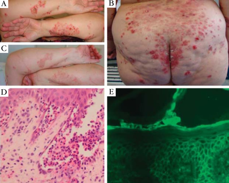
Pemphigus herpetiformis: (A) patient presenting grouped vesicles,
blisters, erosions and crusts onto an erythematous skin in a herpetiform pattern
on her forearms; (B) similar lesions on her buttocks and back;
(C) the same patient 10 days after pulse therapy with
methylprednisolone (1 g/day for 3 days), showing a good clinical response;
(D) histopathological exam of a forearm lesion showing suprabasal
blister containing some acantholytic cels, neutrophils and eosinophils, besides
focal eosinophilic spongiosis (HE 400x); (E) DIF of perilesional skin
showing intercellular distribution of IgG and C3 throughout the entire
epidermis

IgA Pemphigus (IEN type): (A) and (B) vesicles,
blisters, pustules and crusts confluent, occupying almost the entire trunk, neck
and part of the upper limbs; (C) DIF: IgA deposits
intercellular;( D) IIF showing presence of IgA in the patient´s
sera (1:640)

Paraneoplastic pemphigus: (A); ulcer in the side of the tongue, organ typically
affected in paraneoplastic pemphigus. This patient also had erosions in the jugal
mucosa and gingival enanthema. The diagnosis of an abdominal myofibroblastic tumor
led to the suspicion of PNP, which was confirmed by indirect immunofluorescence in
rat bladder and immunoblotting. The patient was initially treated with prednisone
and azathioprine, and later, rituximab, with improvement; (B) DIF of perilesional
patient's skin showing intercellular and basement membrane zone staining (IgG,
10x); (C) IIF in transitional epithelium: positive test for a patient with PNP
(rat bladder, 10x); (D) Immunoblotting (left) and immunoprecipitation (right):
detection of antibodies directed against periplakin (190 kd) and envoplakin (210
kd) is a criterium for diagnosis

Paraneoplastic Pemphigus in patient presenting non-Hodgkin B-cell linfoma: (A)
lesions affecting the lips and oral mucosa; (B) erosions on the back; (C) blisters
on the hands; (D)histopathology showing suprabasal blister containing acantholytic
cells (HE 40x); (E) closer view of the acantholytic cells and loss of
intercellular cohesiveness (HE 400x); (F) DIF showing intercellular deposits of
IgG and C3, and also linear deposits in the BMZ (DIF, 400x); (G) IIF (rat bladder)
showing intercellular distribution of anti-IgG (1:320)
References
-
- Patrício P, Ferreira C, Gomes MM, Filipe P. Autoimmune bullous dermatoses: a review. Ann N Y Acad Sci. 2009 Sep;1173:203–210. - PubMed
-
- Amagai M, Hashimoto T, Green KJ, Shimizu N, Nishikawa T. Antigen-specific immunoadsorption of pathogenic autoantibodies in pemphigus foliaceus. J Invest Dermatol. 1995;104:895–901. - PubMed
-
- Amagai M, Klaus-Kovtun V, Stanley JR. Autoantibodies against a novel epithelial cadherin in pemphigus vulgaris, a disease of cell adhesion. Cell. 1991;67:869–877. - PubMed
-
- Robinson ND, Hashimoto T, Amagai M, Chan LS. The new pemphigus variants. J Am Acad Dermatol. 1999;40:649–671. - PubMed
-
- Floden CH, Centale H. A case of clinically typical dermatitis herpetiformis (M. Duhring) presenting acantholysis. Acta Derm Venereol. 1955;35:128–131. - PubMed
Publication types
MeSH terms
Substances
LinkOut - more resources
Full Text Sources
Other Literature Sources
Medical
Miscellaneous