. 2014 Mar 14;343(6176):1260-3.
doi: 10.1126/science.1248943.

Complement is activated by IgG hexamers assembled at the cell surface

Affiliations

Complement is activated by IgG hexamers assembled at the cell surface

Christoph A Diebolder et al. Science. .

Abstract

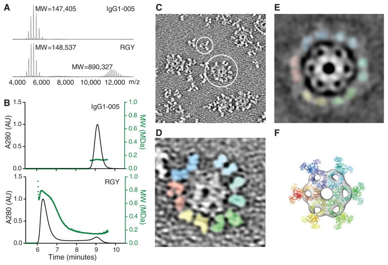
Complement activation by antibodies bound to pathogens, tumors, and self antigens is a critical feature of natural immune defense, a number of disease processes, and immunotherapies. How antibodies activate the complement cascade, however, is poorly understood. We found that specific noncovalent interactions between Fc segments of immunoglobulin G (IgG) antibodies resulted in the formation of ordered antibody hexamers after antigen binding on cells. These hexamers recruited and activated C1, the first component of complement, thereby triggering the complement cascade. The interactions between neighboring Fc segments could be manipulated to block, reconstitute, and enhance complement activation and killing of target cells, using all four human IgG subclasses. We offer a general model for understanding antibody-mediated complement activation and the design of antibody therapeutics with enhanced efficacy.


Figures

Fig. 1
Fig. 1. C1q binding and complement activation by antibody hexamers
(A) IgG hexamer crystal packing of IgG1-b12 (1HZH). The dashed enclosure indicates a single IgG molecule. The C1q binding residue Lys322 is indicated in red. (B) Surface map depicting the Fc-Fc interface. Residues interacting with the Fc-binding peptide DCAWHLGELVWCT are indicated in blue. (C) The Fc-binding peptide inhibits CDC mediated by IgG1-7D8 (Raji cells) and IgG1-005 (Daudi cells). Data are average values ± SD (N = 3); one-way analysis of variance followed by Dunnett’s multiple comparison post hoc test: *P < 0.05, ***P < 0.001. (D) C1q binding to CD20+ Raji cells opsonized with wild-type or mutated CD20 antibody IgG1-7D8. MESF, molecules of equivalent soluble fluorochrome. A representative example is shown (N = 3). (E) CDC of Raji cells opsonized with wild-type and mutated IgG1-7D8. A representative example is shown (N = 3). The absence of CDC without added C1q indicates classical pathway activation. (F and G) CDC of K439E and S440K, abrogated in single point mutants, is restored in an IgG1-7D8 double mutant [(F), Raji cells] and by mixing single mutants of IgG1-7D8 (F) or IgG1-005 [(G), Daudi cells]. Representative examples are shown (N = 3). Amino acid abbreviations: A, Ala; C, Cys; D, Asp; E, Glu; F, Phe; G, Gly; H, His; I, Ile; K, Lys; L, Leu; M, Met; N, Asn; P, Pro; Q, Gln; R, Arg; S, Ser; T, Thr; V, Val; W, Trp; Y, Tyr.
Fig. 2
Fig. 2. Increased CDC by enhanced hexamer formation
(A and B) Increased CDC of Daudi cells by IgG1-7D8-E345R (A) and IgG1-005-E345R (B) relative to wild-type antibodies. Representative examples are shown (N = 4). (C) E345R mutants of IgG1-005 isotype variants induces CDC of Daudi cells more potently than wild-type (WT) IgG2, IgG3, and IgG4; IgGs were tested at 10 μg/ml. Data are average values ± SD (N = 3); two-sided unpaired t test with Welch’s correction: n.s., not significant; **P < 0.01, ***P < 0.001. (D) IgG1-005-RGY induced C4d generation in normal human serum. Data are average values ± SD (N = 3). Two-sided unpaired t test with Welch’s correction; *P < 0.05. Heat-aggregated IgG (HAG) was used as a positive control. (E) IgG1-005-RGY showed enhanced CDC activity of Ramos cells relative to wild-type IgG1-005 and IgG1-005-E345R. A representative example is shown (N = 3).
Fig. 3
Fig. 3. Solution-phase hexamers formed by triple mutant IgG1-005-RGY
(A) Native mass spectrometry of IgG1-005 indicating a molecular weight (MW) of 147,405 daltons and IgG1-005-RGY showing MWs of a monomer (148,537 daltons) and a hexamer (890,327 daltons) (table S3). The hexameric state was confirmed in experiments using different conditions (N = 6); m/z, mass/charge ratio. (B) Overlay of HP-SEC–MALS profiles [absorbance at 280 nm (A280), black, left axis; MW, green, right axis] of IgG1-005 (top) and IgG1-005-RGY (bottom) shows that ~79% IgG1-005-RGY eluted as hexamer and ~21% as monomer, whereas >99% of IgG1-005 eluted as monomer. A representative example is shown (N = 3). (C to F) ET of negatively stained IgG1-005-RGY. (C) ET overview image showing a monomer (small circle) and a hexamer (large circle). (D) Representative hexamer with colored Fab pairs. (E) ET average of 200 subtomograms at a resolution of 2.9 nm. (F) Surface rendering of a symmetrized Fc ring with docked 1HZH hexamer.
Fig. 4
Fig. 4. Visualization of antibody-C1 complexes on antigen-coated liposomes
(A) Subtomogram average of antibody-C1 at >6 nm resolution shown as an isosurface. Heights indicate distances to the membrane center. (B) Vertical section through cryo-ET average. White arrows indicate the positions of sections shown in (C) to (E). (C) Top horizontal section showing four putative C1q globular headpieces. (D) Center section showing a dense hexagonal platform. (E) Bottom section showing six putative antigen-binding Fab arms. (F) Side view of the IgG1-b12–based hexamer model placed into the six-fold symmetrized density of the lower cryo-ET platform (top) and hexamer model with docked C1q headpieces (bottom). (G) CDC of CD20+ Raji cells by (functionally monovalent) bispecific antibody IgG1-7D8/b12. A representative example is shown (N = 3). (H) CDC of EGFR+ A431 cells by (functionally monovalent) bispecific antibody IgG1-2F8/b12. A representative example is shown (N = 4). IgG1-b12 against HIV-1 gp120 contributed the innocuous Fab arm.

References

    1. Ricklin D, Hajishengallis G, Yang K, Lambris JD. Nat Immunol. 2010;11:785–797. - PMC - PubMed
    1. Dunkelberger JR, Song WC. Cell Res. 2010;20:34–50. - PubMed
    1. Gaboriaud C, et al. Trends Immunol. 2004;25:368–373. - PubMed
    1. Kishore U, Reid KB. Immunopharmacology. 2000;49:159–170. - PubMed
    1. Hughes-Jones NC, Gardner B. Mol Immunol. 1979;16:697–701. - PubMed

Publication types