. 2014 May;7(5):547-59.
doi: 10.1242/dmm.014068. Epub 2014 Mar 13.

Complexes of Usher proteins preassemble at the endoplasmic reticulum and are required for trafficking and ER homeostasis

Affiliations

Complexes of Usher proteins preassemble at the endoplasmic reticulum and are required for trafficking and ER homeostasis

Bernardo Blanco-Sánchez et al. Dis Model Mech. 2014 May.

Abstract

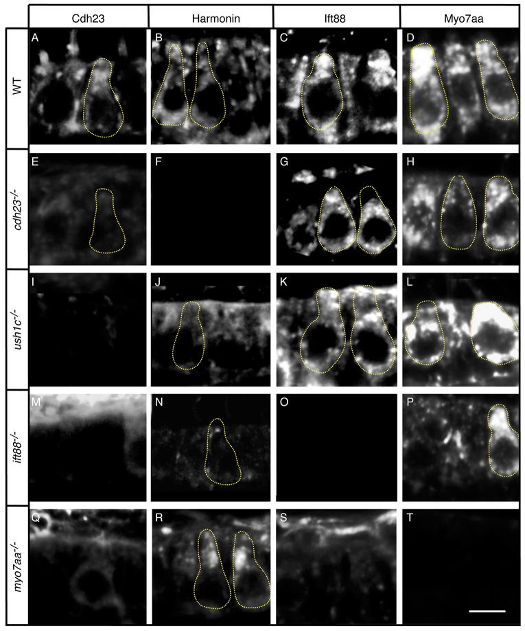
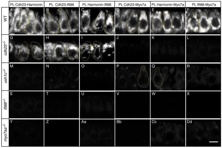
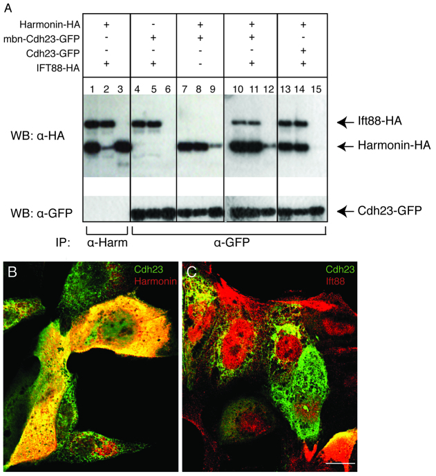
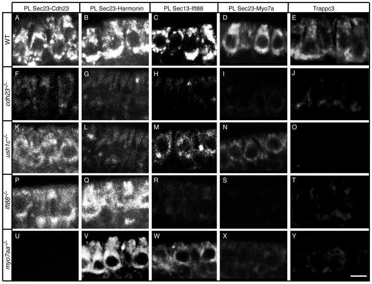
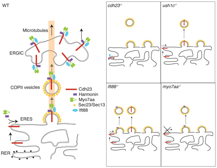
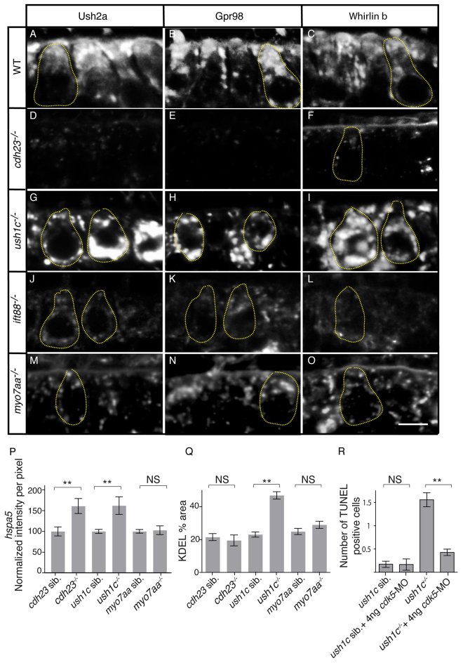
Usher syndrome (USH), the leading cause of hereditary combined hearing and vision loss, is characterized by sensorineural deafness and progressive retinal degeneration. Mutations in several different genes produce USH, but the proximal cause of sensory cell death remains mysterious. We adapted a proximity ligation assay to analyze associations among three of the USH proteins, Cdh23, Harmonin and Myo7aa, and the microtubule-based transporter Ift88 in zebrafish inner ear mechanosensory hair cells. We found that the proteins are in close enough proximity to form complexes and that these complexes preassemble at the endoplasmic reticulum (ER). Defects in any one of the three USH proteins disrupt formation and trafficking of the complex and result in diminished levels of the other proteins, generalized trafficking defects and ER stress that triggers apoptosis. ER stress, thus, contributes to sensory hair cell loss and provides a new target to explore for protective therapies for USH.

Keywords: Cadherin23; ER stress; Hair cell; Harmonin; Ift88; Myo7aa; Trafficking; Usher syndrome; Zebrafish.


Figures

Fig. 1
Fig. 1
Zebrafish cdh23, ush1c, myo7aa and ift88 mutants have mechanoreceptor structural defects. (A–E) Confocal projections of anterior maculae labeled with phalloidin in (A) wild-type (WT) sibling, (B) cdh23tj264a, (C) ush1cfh293, (D) ift88tz288b and (E) myo7aaty220d mutants. (F–H) Confocal optical sections of double immunolabeling of (F) Harmonin (green) and Ift88 (red), (G) Harmonin (green) and Cdh23 (red), and (H) Harmonin (green) and Myo7aa (red). Individual hair cells are shown. (I) Quantification of total number of hair bundles. n≥6 analyzed anterior maculae. (J) Quantification of total number of hair cells. n≥5 analyzed anterior maculae. (K) Quantification of hair cell death in mutants and wild-type siblings. Number: average number of TUNEL-positive hair cells per macula. n≥16 analyzed anterior maculae. Student’s t-test: **P<0.01, *P<0.05. NS, non significant. Black lines represent ± s.e.m. Scale bars: 5 μm.
Fig. 2
Fig. 2
Cdh23 and Harmonin require Ift88 and Myo7aa for normal localization in hair cells. Immunolabeling in (A–D) wild-type siblings or (E–H) cdh23, (I–L) ush1c, (M–P) ift88 and (Q–T) myo7aa mutants of (A,E,I,M,Q) Cdh23, (B,F,J,N,R) Harmonin, (C,G,K,O,S) Ift88 and (D,H,L,P,T) Myo7aa. Confocal views of anterior macula hair cells are shown. Confocal sections are 0.8 μm thick, whereas the neck and basal region of the hair cell have an estimated diameter of 2–2.5 μm and 5–6 μm, respectively. Thus, not all confocal sections contain a hair bundle, and the observed shape of the hair bundle varies according to the mounting angle (see also supplementary material Fig. S3). Some individual hair cells are outlined by dotted lines. WT, wild type. Scale bar: 5 μm.
Fig. 3
Fig. 3
Cdh23, Harmonin, Ift88 and Myo7aa proteins are in close proximity. Proximity labeling (PL) of (A,G,M,S,Y) Cdh23 and Harmonin, (B,H,N,T,Z) Cdh23 and Ift88, (C,I,O,U,Aa) Harmonin and Ift88, (D,J,P,V,Bb) Cdh23 and Myo7a, (E,K,Q,W,Cc) Harmonin and Myo7a, and (F,L,R,X,Dd) Ift88 and Myo7. (A–F) Wild-type (WT) siblings; (G–L) cdh23, (M–R) ush1c, (S–X) ift88 and (Y-Dd) myo7aa mutants. Confocal views of anterior macula hair cells. Some individual hair cells are outlined by dotted lines. Scale bar: 5 μm.
Fig. 4
Fig. 4
Harmonin and Cdh23 bind directly to each other, but not to Ift88. (A) Co-immunoprecipitation. MDCK cells were transfected as indicated by (+) with combinations of DNA vectors encoding Harmonin-HA, mbn-Cdh23-GFP, Cdh23-GFP or IFT88-HA. Input lanes: 1,4,7,10,13. Unbound fraction lanes: 2,5,8,11,14. Bound fraction lanes: 3,6,9,12,15. Immunoprecipitation was performed with an antibody against Harmonin (α-Harm) or GFP (α-GFP). (B,C) Confocal sections of transfected MDCK cells. (B) Double immunolabeling of mbn-Cdh23-GFP (green) and Harmonin (red). (C) Double immunolabeling of mbn-Cdh23-GFP (green) and Ift88 (red). Harmonin-HA: full-length zebrafish Harmonin isoform A fused to HA-tag at the C-terminal. mbn-Cdh23-GFP: zebrafish Cdh23 membrane bound cytoplasmic domain fused to GFP at the C-terminal. Cdh23-GFP: zebrafish Cdh23 cytoplasmic domain fused to GFP at the C-terminal. Ift88-HA: full-length zebrafish Ift88 fused to HA-tag at the C-terminal. Scale bar: 7.5 μm.
Fig. 5
Fig. 5
The Cdh23, Harmonin, Ift88 and Myo7aa protein complex is present at the ER and associated vesicles. Proximity labeling (PL) of (A–D) wild-type (WT) siblings, and (F–I) cdh23, (K–N) ush1c, (P–S) ift88 and (U–X) myo7aa mutants. Proximity labeling of (A,F,K,P,U) Sec23 and Cdh23, (B,G,L,Q,V) Sec23 and Harmonin, (C,H,M,R,W) Sec13 and Ift88, and (D,I,N,S,X) Sec23 and Myo7a (recognizes Myo7aa and possibly Myo7ab; see Materials and Methods). (E,J,O,T,Y) Immunolabeling of Trappc3 in (E) WT, and (J) cdh23, (O) ush1c, (T) ift88 and (Y) myo7aa mutants. Confocal views of anterior macula hair cells. Scale bar: A–D,F,I,K–N,P–S,U–X: 7.5 μm; E,J,O,T,Y: 5 μm.
Fig. 6
Fig. 6
The Cdh23, Harmonin, Ift88 and Myo7aa protein complex preassembles at the ER. Model for assembly and trafficking of USH proteins. Under normal conditions in wild-type (WT) animals, Cadherin23, Harmonin, Ift88 and Myo7aa preassemble into a complex at the level of the ER. Interaction between Cdh23 and Myo7aa is required for Cdh23 translocation to the ERES. All four proteins are required for assembly of a functional complex. Once assembled, the complex translocates to ER exit sites (ERES), engages components of the COPII system (Sec23 and Sec13), and enters the secretory pathway where it is trafficked to the ER-Golgi intermediate complex (ERGIC) en route to final destinations. Loss-of-function mutations in cdh23, ush1c, ift88 and myo7aa disrupt assembly of the complex in various ways. RER, rough endoplasmic reticulum.
Fig. 7
Fig. 7
The Cdh23, Harmonin and Myo7aa protein complex is required for proper trafficking of USH2 proteins and can trigger ER stress when defective. (A–O) Immunolabeling of USH2 proteins in (A–C) wild-type (WT) siblings, and (D–F) cdh23, (G–I) ush1c, (J–L) ift88 and (M–O) myo7aa mutants with antibodies against Ush2a (A,D,G,J,M), Gpr98 (B,E,H,K,N) and Whirlinb (C,F,I,L,O). (P) Levels of hspa5 expression based on in situ hybridization technique in whole-mount larvae. For ush1c siblings n=57; ush1c−/− n=51; cdh23 siblings n=35; for cdh23−/− n=33; myo7aa siblings n=24; for myo7aa−/− n=24 analyzed anterior maculae. (Q) Quantification of percentage area per hair cell containing KDEL expression. For ush1c siblings n=85; ush1c−/− n=125; cdh23 siblings n=66; for cdh23−/− n=60; myo7aa siblings n=60; myo7aa−/− n=54 analyzed hair cells for each genotype. (R) Knockdown of cdk5 rescues ER-stress-induced cell death in the ush1c mutant. Bars show average number of TUNEL-positive cells. For ush1c siblings n=52; ush1c siblings + 4 ng cdk5-MO n=18; ush1c−/− n=70; ush1c−/− + 4 ng cdk5-MO n=70 analyzed anterior maculae. Average ± s.d. Statistics were conducted with Student’s t-test. **P<0.01. NS, non significant. Some individual hair cells are outlined by dotted lines. MO, morpholino. Scale bar: 5 μm.

References

    1. Adato A., Lefèvre G., Delprat B., Michel V., Michalski N., Chardenoux S., Weil D., El-Amraoui A., Petit C. (2005a). Usherin, the defective protein in Usher syndrome type IIA, is likely to be a component of interstereocilia ankle links in the inner ear sensory cells. Hum. Mol. Genet. 14, 3921–3932 - PubMed
    1. Adato A., Michel V., Kikkawa Y., Reiners J., Alagramam K. N., Weil D., Yonekawa H., Wolfrum U., El-Amraoui A., Petit C. (2005b). Interactions in the network of Usher syndrome type 1 proteins. Hum. Mol. Genet. 14, 347–356 - PubMed
    1. Bahloul A., Michel V., Hardelin J. P., Nouaille S., Hoos S., Houdusse A., England P., Petit C. (2010). Cadherin-23, myosin VIIa and harmonin, encoded by Usher syndrome type I genes, form a ternary complex and interact with membrane phospholipids. Hum. Mol. Genet. 19, 3557–3565 - PMC - PubMed
    1. Blasic J. R., Jr, Brown R. L., Robinson P. R. (2012). Phosphorylation of mouse melanopsin by protein kinase A. PLoS ONE 7, e45387. - PMC - PubMed
    1. Boëda B., El-Amraoui A., Bahloul A., Goodyear R., Daviet L., Blanchard S., Perfettini I., Fath K. R., Shorte S., Reiners J., et al. (2002). Myosin VIIa, harmonin and cadherin 23, three Usher I gene products that cooperate to shape the sensory hair cell bundle. EMBO J. 21, 6689–6699 - PMC - PubMed

Publication types

MeSH terms

Substances