. 2014 May 2;289(18):12245-63.
doi: 10.1074/jbc.M114.556639. Epub 2014 Mar 13.

Glycosylphosphatidylinositol anchoring directs the assembly of Sup35NM protein into non-fibrillar, membrane-bound aggregates

Affiliations

Glycosylphosphatidylinositol anchoring directs the assembly of Sup35NM protein into non-fibrillar, membrane-bound aggregates

Karen E Marshall et al. J Biol Chem. .

Abstract

In prion-infected hosts, PrPSc usually accumulates as non-fibrillar, membrane-bound aggregates. Glycosylphosphatidylinositol (GPI) anchor-directed membrane association appears to be an important factor controlling the biophysical properties of PrPSc aggregates. To determine whether GPI anchoring can similarly modulate the assembly of other amyloid-forming proteins, neuronal cell lines were generated that expressed a GPI-anchored form of a model amyloidogenic protein, the NM domain of the yeast prion protein Sup35 (Sup35(GPI)). We recently reported that GPI anchoring facilitated the induction of Sup35(GPI) prions in this system. Here, we report the ultrastructural characterization of self-propagating Sup35(GPI) aggregates of either spontaneous or induced origin. Like membrane-bound PrPSc, Sup35(GPI) aggregates resisted release from cells treated with phosphatidylinositol-specific phospholipase C. Sup35(GPI) aggregates of spontaneous origin were detergent-insoluble, protease-resistant, and self-propagating, in a manner similar to that reported for recombinant Sup35NM amyloid fibrils and induced Sup35(GPI) aggregates. However, GPI-anchored Sup35 aggregates were not stained with amyloid-binding dyes, such as Thioflavin T. This was consistent with ultrastructural analyses, which showed that the aggregates corresponded to dense cell surface accumulations of membrane vesicle-like structures and were not fibrillar. Together, these results showed that GPI anchoring directs the assembly of Sup35NM into non-fibrillar, membrane-bound aggregates that resemble PrPSc, raising the possibility that GPI anchor-dependent modulation of protein aggregation might occur with other amyloidogenic proteins. This may contribute to differences in pathogenesis and pathology between prion diseases, which uniquely involve aggregation of a GPI-anchored protein, versus other protein misfolding diseases.

Keywords: Amyloid; Electron Microscopy (EM); Glycosylphosphatidylinositol Anchors; Prions; Protein Aggregation; Protein Misfolding; Protein Self-assembly.


Figures

FIGURE 1.
FIGURE 1.
GPI-anchored Sup35 and control constructs. The amyloidogenic NM domain of the yeast prion Sup35 was expressed as a fusion to a GPI-anchored fluorescent protein (GFP or mCherry) in mouse N2a neuroblastoma cells. GPI anchorless constructs were generated by insertion of a stop codon immediately prior to the GPI anchor addition sequence. An N-terminal signal peptide (SP) sequence was included in all constructs, which directs the proteins to the secretory pathway in mammalian cells. Shaded segment, Myc (GFP constructs) or HA (mCherry constructs) antibody epitope tag.
FIGURE 2.
FIGURE 2.
Stably transfected cell lines produce aggregated or non-aggregated forms of Sup35-GFPGPI. Cloned N2a cell lines stably expressing the Sup35-GFPGPI construct propagated either aggregated (Agg) (C) or non-aggregated (Sol) (B) forms of Sup35-GFPGPI. Aggregate-positive cells (C) express high levels of Sup35-GFPGPI, with aggregates appearing as areas of bright, punctate fluorescence. The higher magnification inset highlights the two types of aggregate morphology observed: irregular (arrowhead) and contoured (arrow). The distribution of Sup35-GFPGPI-Sol (B) resembles that of the GFPGPI control construct-expressing cells (A), with no aggregates observed. Samples were imaged by wide field fluorescence microscopy. Scale bar, 40 μm; inset scale, 20 μm.
FIGURE 3.
FIGURE 3.
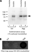
Sup35-GFPGPI aggregates are detergent-insoluble. Sup35-GFPGPI-Sol and -Agg cell lysates were assayed for detergent-insoluble Sup35-GFPGPI by two independent methods. Lysates were normalized for total protein prior to assay. A, immunoblot probed with anti-Sup35 N domain antibody following sedimentation assay with 1% sarkosyl. The arrow near 80 kDa corresponds to Sup35NM. B, dot blot probed with anti-GFP antibody following filter trap assay with 4% SDS. Detergent-insoluble material was only found in cells expressing aggregated forms of Sup35-GFPGPI.
FIGURE 4.
FIGURE 4.
Sup35-GFPGPI aggregates are resistant to digestion with chymotrypsin. Sup35-GFPGPI-Sol (lanes 1–6) and Sup35-GFPGPI-Agg (lanes 7–12) cell lysates were digested with increasing concentrations of chymotrypsin (Ct) and analyzed by immunoblot, probing with anti-Sup35 N domain antibody. Arrow, Sup35-GFPGPI. Chymotrypsin-resistant material was only observed in Sup35-GFPGPI-Agg cell lysates, suggesting a conformational change in Sup35NM.
FIGURE 5.
FIGURE 5.
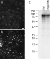
Aggregate formation in Sup35-GFPGPI cells is not related to expression level. A and B, wide field fluorescence microscopy images of Sup35-GFPGPI-Sol (A) and Sup35-GFPGPI-Agg (clone D8) cells. Clone D8 was an independent clone in which aggregates rose spontaneously. As shown by quantitative immunoblotting (C) the overall expression level of Sup35-GFPGPI was 52 ± 3.9% (n = 4) lower in Sup35-GFPGPI-Agg (D8) cells than in Sup35-GFPGPI-Sol cells that did not contain any aggregates. Scale bar, 20 μm.
FIGURE 6.
FIGURE 6.
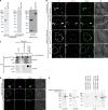
Aggregates from Sup35-GFPGPI-Agg cells seed formation of GPI anchorless Sup35-mC aggregates. A, stably transfected cells expressing anchorless forms of mC or Sup35-mC were assayed by immunoblot for expression of the respective proteins. Cell lysates were normalized for total protein and probed with anti-HA or anti-Sup35 M domain antibodies. Culture supernatants were assayed for mCherry-tagged protein by immunoprecipitation. High levels of anchorless mC and Sup35-mC were released to culture supernatants. B, in situ seeding by fixed Sup35-GFPGPI aggregates. Cells were chemically fixed prior to incubation with culture supernatants from cells expressing the indicated forms of GPI-anchorless mCherry protein and confocal imaging. Arrows, Sup35-GFPGPI aggregates. Arrowheads, newly induced Sup35-mC aggregates. White in the merge panel (G) indicates areas of co-localization. Images correspond to a single 0.5-μm optical z slice from near the middle of the cells. Scale bar, 10 μm. C, cell-free seeding assay. Microsome fractions from Sup35-GFPGPI-Agg cells as a source of seeds or GFPGPI control cells (or PBS buffer control) were incubated with the indicated substrates. Reactions were analyzed by filter trap assay to detect either preexisting (anti-GFP panels) or newly induced mCherry-tagged (anti-RFP panels; anti-RFP binds mCherry) SDS-insoluble aggregates. D, in situ seeding by Sup35-GFPGPI aggregates on live cells. Live cells were incubated with culture supernatants from cells expressing Sup35-mC followed by confocal imaging. Arrows, Sup35-GFPGPI aggregates. Arrowhead, newly induced Sup35-mC aggregate. White in the merge panel (C) indicates the area of co-localization. Images correspond to a single 0.5-μm optical z slice from near the middle of the cells. Scale bar, 10 μm. E, chymotrypsin resistance assay. After imaging, cultures in D were lysed, digested with chymotrypsin, and immunoblotted with anti-HA tag antibody (lanes 5–10). Control samples of Sup35-mC culture supernatant treated in parallel were also immunoblotted with anti-Sup35 N (lanes 1 and 2) and M domain antibodies (lanes 3 and 4). Chymotrypsin-negative lanes contain one-quarter sample equivalents loaded in chymotrypsin-treated lanes. Arrow, full-length Sup35-mC. Open arrowhead, chymotrypsin-truncated Sup35-mC.
FIGURE 7.
FIGURE 7.
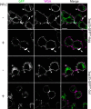
Cell surface Sup35-GFPGPI aggregates are resistant to release by PIPLC. Distribution of aggregated (Sup35-GFPGPI-Agg; rows 1 and 2) and non-aggregated (Sup35-GFPGPI-Sol; rows 3 and 4) Sup35-GFPGPI in cells with and without PIPLC treatment is shown. Plasma membrane staining at 4 °C with WGA (magenta channel) was used as a marker for the cell surface. Areas of WGA and Sup35-GFPGPI co-localization appear white in the merge panels. The arrowheads indicate non-aggregated Sup35-GFPGPI in both soluble and aggregated cell cultures. This staining was absent in samples treated with PIPLC, indicating removal from the cell surface. Aggregates (arrows) were resistant to PIPLC-induced release (row 2, arrows). Samples were imaged by confocal microscopy, and images represent a single 0.5-μm optical z slice. Scale bar, 10 μm.
FIGURE 8.
FIGURE 8.
Sup35-mCGPI aggregates do not stain with Thioflavin T. Cultures were stained with the amyloid-specific dye Thioflavin T. mCGPI, negative control cells stably expressing GPI-anchored mCherry (A–C). mCGPI + rSup35NM fibrils, mCGPI cells treated with rSup35NM amyloid fibrils as a positive control (D–F). Sup35-mCGPI, cells stably expressing high levels of aggregated Sup35-mCGPI (G–I). Sup35-mCGPI was used instead of GFP-tagged versions of the construct for these experiments due to spectral overlap between GFP and ThT. Sup35-mCGPI aggregates did not exhibit any fluorescence when treated with ThT (G–I, arrows), unlike rSup35NM fibrils (D, arrowheads). Scale bar, 10 μm.
FIGURE 9.
FIGURE 9.
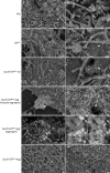
Ultrastructure of membrane-bound Sup35-GFPGPI aggregates by scanning electron microscopy. Untransfected N2a cells (N2a) (A and B), cells expressing a control GFPGPI construct (GFPGPI) (C and D), Sup35-GFPGPI-Sol (E and F), and Sup35-GFPGPI-Agg (G–M) cells were immunogold-labeled without permeabilization using anti-GFP antibody and imaged by SEM. J and K, higher magnification images of the dashed area in I. Negative control immunogold-labeled samples of Sup35-GFPGPI-Agg cells where primary (L) or secondary (M) antibody was omitted are also shown. With the exception of K, all images contain a mix of backscattered and secondary electron signals (BSE/SE mix), and thus gold particles appear as white spots. K shows only the secondary electron (SE) signal corresponding to the same field of view in J to provide a clear, high resolution image of gold-labeled structures. Densely labeled areas (arrows) correspond to aggregates. More diffuse labeling was observed in cells transfected with the GFPGPI control construct and Sup35-GFPGPI-Sol cells (arrowheads). No fibrillar structures were observed on Sup35-GFPGPI-Agg cells. Scale bars, 600 nm (A, C, E, G, L, and M), 200 nm (B, D, F, and H), 120 nm (I), and 40 nm (J and K).
FIGURE 10.
FIGURE 10.
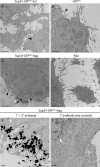
Ultrastructure of membrane-bound Sup35-GFPGPI aggregates by transmission electron microscopy. Cells were immunogold-labeled without permeabilization with anti-GFP antibody and imaged by TEM. The enhanced FluoroNanogold particles were observed as large, electron-dense particles (black arrows and arrowheads). White arrows in E and F indicate very small electron-dense objects that corresponded to background from the gold enhancement procedure to detect nanogold, as shown by their appearance in negative control samples where the FluoroNanogold secondary was omitted (e.g. F). Scale bars, 2 μm (A–D) and 100 nm (E and F). Dense accumulations of gold particles were only present in Sup35-GFPGPI-Agg cells (C and E, black arrowheads) and did not appear fibrillar.
FIGURE 11.
FIGURE 11.
Imaging FluoroNanogold-labeled samples by wide field fluorescence microscopy. Aggregates showed strong GFP fluorescence (GFP channel, arrowheads) with proportionally dimmer fluorescence of cell membranes in areas without aggregates. Aggregates were strongly immunolabeled as shown in the Alexa Fluor 594-FluoroNanogold channel (arrowheads) and white regions in the merge panels. Strong fluorescent labeling correlates with areas of dense gold deposition in EM images. G–I, higher magnification images of aggregates indicated in D–F. Scale bars, 20 μm.
FIGURE 12.
FIGURE 12.
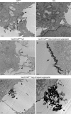
Immunoperoxidase TEM images of Sup35-GFPGPI aggregates. Control cells (A and B) and cells producing non-aggregated (C) or aggregated (D–F) Sup35-GFPGPI were stained without permeabilization with anti-GFP primary antibody and secondary antibody conjugated to HRP, which catalyzes the conversion of DAB substrate into a product detectable by TEM. Staining was present all along the surface membranes of transfected cells, with occasional small regions of more intense staining in both GFPGPI and Sup35-GFPGPI-Sol cells (arrowheads). The small intensely stained regions probably correspond to concentrations of GPI-anchored protein in individual raft membrane microdomains. Sup35-GFPGPI-Agg cells showed a similar staining pattern but were distinguished by the presence of very large, intensely stained regions that appeared to consist of cell surface accumulations of vesicles (arrows) resembling aggregate structures observed by immunogold labeling. The location, morphology, relative staining intensity, and specificity to Sup35-GFPGPI-Agg cells all strongly indicate that these large, intensely stained regions correspond to Sup35-GFPGPI aggregates. No staining was observed on N2a control cells (B) or on additional control samples for all cells where incubation with primary antibody was omitted (data not shown). All scale bars, 500 nm.
FIGURE 13.
FIGURE 13.
Visualization of biotinylated rSup35NM fibrils added to GFPGPI control cells. A, immunoblot of biotinylated rSup35NM fibrils with anti-Sup35M antibody (α-Sup35M) detected both SDS-resistant oligomeric (bracket) and monomeric (arrow) species that corresponded to biotinylated bands detected with streptavidin in a parallel immunoblot (Biotin). Unlabeled non-aggregated rSup35NM (Non-aggregated) is shown as a control. Two dilutions of each sample were loaded. Vertical white lines indicate removal of irrelevant lanes. B, TEM of negatively stained biotinylated rSup35NM fibrils (scale bar, 100 nm). C, wide field fluorescence microscopy images of GFPGPI cells treated with biotinylated fibrils and immunolabeled using FluoroNanogold. For cells treated with fibrils, the immunostaining (SA-FNG; middle panel) appeared as both punctate spots (arrows) and more uniform cell surface labeling (arrowhead). Negative control samples using either GFPGPI cells not treated with fibrils and labeled (left column) or GFPGPI cells treated with fibrils and labeled with streptavidin-conjugated FluoroNanogold pre-adsorbed with biotin (right column) demonstrate labeling specificity. SA-FNG, FluoroNanogold channel. GFP, GFP channel. Scale bars, 10 μm. D, TEM visualization of biotinylated rSup35NM fibrils added to GFPGPI cells. GFPGPI cells were treated with fibrils and immunolabeled as in C. Fibrils (black arrowheads) were clearly visible in both FluoroNanogold-labeled (subpanel B) and unlabeled (subpanel C) samples. No fibrils or immunolabeling were observed in untreated cells (subpanel A). Sup35-GFPGPI aggregates (arrows) on Sup35-GFPGPI-Agg cells immunolabeled with anti-GFP (subpanels D and E) or without primary antibody (subpanel F) and processed using the same modified gold enhancement procedure as in subpanels A–C are shown for comparison. Note that subpanels E and F were acquired at twice the magnification of subpanels A–D and that immunolabeled structures are not fibrillar. Scale bars, 100 nm.
FIGURE 14.
FIGURE 14.
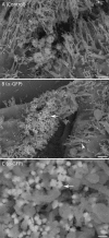
Ultrastructure of rSup35NM amyloid fibril-induced Sup35-GFPGPI aggregates by SEM. Aggregates induced in Sup35-GFPGPI-Sol cells following treatment with rSup35NM amyloid fibrils are shown 41 passes post-fibril treatment. Non-permeabilized cells were stained with anti-GFP mouse monoclonal primary antibody (α-GFP) or an irrelevant mouse monoclonal primary antibody (3F4) as a negative control (control) and FluoroNanogold secondary antibody. Gold particles appear as bright spots. Fibril-induced aggregates looked similar to those that arose spontaneously in Sup35-GFPGPI-Agg cells. Scale bars, 800 nm (A and B) and 100 nm (C).

References

    1. Caughey B. W., Dong A., Bhat K. S., Ernst D., Hayes S. F., Caughey W. S. (1991) Secondary structure analysis of the scrapie-associated protein PrP 27–30 in water by infrared spectroscopy. Biochemistry 30, 7672–7680 - PubMed
    1. Pan K.-M., Baldwin M., Nguyen J., Gasset M., Serban A., Groth D., Mehlhorn I., Huang Z., Fletterick R. J., Cohen F. E., Prusiner S. B. (1993) Conversion of α-helices into β-sheets features in the formation of the scrapie prion protein. Proc. Natl. Acad. Sci. U.S.A. 90, 10962–10966 - PMC - PubMed
    1. Prusiner S. B., Groth D. F., Bolton D. C., Kent S. B., Hood L. E. (1984) Purification and structural studies of a major scrapie prion protein. Cell 38, 127–134 - PubMed
    1. Soto C. (2011) Prion hypothesis: the end of the controversy? Trends Biochem. Sci. 36, 151–158 - PMC - PubMed
    1. Jarrett J. T., Lansbury P. T., Jr. (1993) Seeding “One-Dimensional Crystallization” of Amyloid: a Pathogenic Mechanism in Alzheimer's disease and scrapie? Cell 73, 1055–1058 - PubMed

Publication types

MeSH terms

LinkOut - more resources