Eagle's syndrome: a case report
- PMID: 24627843
- PMCID: PMC3949492
- DOI: 10.5125/jkaoms.2014.40.1.43
Eagle's syndrome: a case report
Abstract
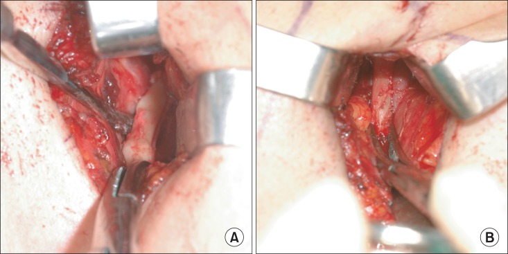
Eagle's syndrome is a disease caused by an elongated styloid process or calcified stylohyoid ligament. Eagle defined the disorder in 1937 by describing clinical findings related to an elongated styloid process, which is one of the numerous causes of pain in the craniofacial and cervical region. The prevalence of individuals with this anatomic abnormality in the adult population is estimated to be 4% with 0.16% of these individuals reported to be symptomatic. Eagle's syndrome is usually characterized by neck, throat, or ear pain; pharyngeal foreign body sensation; dysphagia; pain upon head movement; and headache. The diagnosis of Eagle's syndrome must be made in association with data from the clinical history, physical examination, and imaging studies. Patients with increased symptom severity require surgical excision of the styloid process, which can be performed through an intraoral or an extraoral approach. Here, we report a rare case of stylohyoid ligament bilaterally elongated to more than 60 mm in a 51-year-old female. We did a surgery by extraoral approach and patient's symptom was improved.
Keywords: Eagle syndrome; Elongated styloid process.
Conflict of interest statement
No potential conflict of interest relevant to this article was reported.
Figures


References
-
- Eagle WW. Elongated styloid processes: report of two cases. Arch Otolaryngol. 1937;25:584–587.
-
- Dunn-Ryznyk LR, Kelly CW. Eagle syndrome: a rare cause of dysphagia and head and neck pain. JAAPA. 2010;23:28, 31–32, 48. - PubMed
-
- Yavuz H, Caylakli F, Erkan AN, Ozluoglu LN. Modified intraoral approach for removal of an elongated styloid process. J Otolaryngol Head Neck Surg. 2011;40:86–90. - PubMed
-
- Colby CC, Del Gaudio JM. Stylohyoid complex syndrome: a new diagnostic classification. Arch Otolaryngol Head Neck Surg. 2011;137:248–252. - PubMed
-
- Johnson GM, Rosdy NM, Horton SJ. Manual therapy assessment findings in patients diagnosed with Eagle's Syndrome: a case series. Man Ther. 2011;16:199–202. - PubMed
Publication types
LinkOut - more resources
Full Text Sources
Other Literature Sources