. 2014 Jul;142(3):492-505.
doi: 10.1111/imm.12281.

Disruption of HLA class II antigen presentation in Burkitt lymphoma: implication of a 47,000 MW acid labile protein in CD4+ T-cell recognition

Affiliations

Disruption of HLA class II antigen presentation in Burkitt lymphoma: implication of a 47,000 MW acid labile protein in CD4+ T-cell recognition

Jason M God et al. Immunology. 2014 Jul.

Abstract

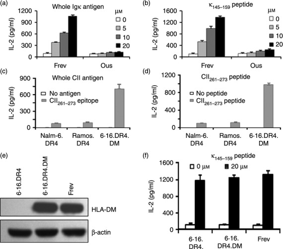
While Burkitt lymphoma (BL) has a well-known defect in HLA class I-mediated antigen presentation, the exact role of BL-associated HLA class II in generating a poor CD4(+) T-cell response remains unresolved. Here, we found that BL cells are deficient in their ability to optimally stimulate CD4(+) T cells via the HLA class II pathway. This defect in CD4(+) T-cell recognition was not associated with low levels of co-stimulatory molecules on BL cells, as addition of external co-stimulation failed to elicit CD4(+) T-cell activation by BL. Further, the defect was not caused by faulty antigen/class II interaction, because antigenic peptides bound with measurable affinity to BL-associated class II molecules. Interestingly, functional class II-peptide complexes were formed at acidic pH 5·5, which restored immune recognition. Acidic buffer (pH 5·5) eluate from BL cells contained molecules that impaired class II-mediated antigen presentation and CD4(+) T-cell recognition. Biochemical analysis showed that these molecules were greater than 30,000 molecular weight in size, and proteinaceous in nature. In addition, BL was found to have decreased expression of a 47,000 molecular weight enolase-like molecule that enhances class II-mediated antigen presentation in B cells, macrophages and dendritic cells, but not in BL cells. These findings demonstrate that BL likely has multiple defects in HLA class II-mediated antigen presentation and immune recognition, which may be exploited for future immunotherapies.

Keywords: Burkitt lymphoma; HLA class II; antigen presentation; enolase-like molecules; immune escape.


Figures

Figure 1
Figure 1
Burkitt lymphoma (BL) and BL type cells are deficient in HLA class II-mediated antigen presentation and CD4+ T-cell recognition. (a) BL type cells (Nalm-6), BL (Ramos), and the B-lymphoblastoid cells (B-LCL) (6.16.DM) were retrovirally transfected to constitutively express a common allele of HLA-DR4 (DR4B*0401). Cells were stained with a pan-HLA-class II antibody (L-243, dotted line) and an HLA-DR4-specific antibody (359-F10, solid line). FACS plots are shown after retroviral transfection. Data are representative of at least three separate experiments. (b) Western blot analysis for HLA-DM proteins in Nalm-6.DR4, Ramos.DR4, and 6.16.DR4.DM cells. (c) Nalm-6.DR4, Ramos.DR4, and 6.16.DR4.DM cell lines were incubated with the synthetic κ145–159 peptide (0–20 μm) for 4 hr, washed, and co-cultured with the κ peptide-specific T-cell hybridoma (1.21) for 24 hr. (d) Cells were incubated with the synthetic κ188–203 peptide (20 μm) for 4 hr, washed and co-cultured with the peptide-specific T-cell hybridoma (2.18a) for 24 hr. (e) Cells were also incubated with the whole antigen IgG κ for overnight, washed, and co-cultured with the κ epitopes-specific T-cell hybridomas (2.18a for κ188–203, 1.21 for κ145–159) for 24 hr. Following incubation, T-cell production of IL-2 in the cell-free culture supernatant was measured by ELISA. Data are expressed as the mean ± SEM of triplicate experiments.
Figure 2
Figure 2
Disruption of class II–antigen presentation and CD4+ T-cell recognition of Burkitt lymphoma (BL). (a) A wild-type B-lymphoblastoid cell line (B-LCL) (Frev) and a wild-type BL line (Ous), which constitutively express HLA-DR4 (DR4B*0401) allele, were incubated with the whole antigen IgG κ (0–20 μm) for overnight. (b) Cells were also incubated with the synthetic κ145–159 peptide (0–20 μm) for 4 hr. After incubation, cells were washed and co-cultured with the κ145–159-specific T-cell hybridoma line (1.21) for 24 hr. (c) Cells were incubated with another self antigen type II collagen (CII; whole CII, 40 μg/ml) for overnight. (d) Cells were also incubated with the synthetic CII261–273 peptide (20 μm) for 4 hr. Cells were then washed and co-cultured with the CII261–273-specific T-cell hybridoma line 4027/99 for 24 hr. The production of IL-2 in the cell-free culture supernatant was quantified by ELISA. (e) Western blot analysis for HLA-DM proteins in 6.16.DR4, 6.16.DR4.DM, and Frev cells. (f) 6.16.DR4, 6.16.DR4.DM and Frev cells were incubated with the synthetic κ145–159 peptide (20 μm) for 4 hr. Cells were washed and co-cultured with the κ145–159-specific T-cell hybridoma line (1.21) for 24 hr. The T-cell production of IL-2 was quantified as described. Data are representative of the mean ± SEM of triplicate experiments.
Figure 3
Figure 3
Analysis of major class II pathway components, requirement of co-stimulation and production of inhibitory cytokines in Burkitt lymphoma (BL)/B-lymphoblastoid cells (B-LCL). (a) RT-PCR analyses of Nalm-6.DR4, Ramos.DR4, and Frev cells for HLA-DM, HLA-DO, and Ii gene expression. β-Actin was used as a loading control. Data are representative of at least three separate experiments. (b) Co-stimulation does not overcome the BL defect in HLA class II antigen presentation. Nalm-6.DR4, Ramos.DR4, and 6.16.DR4.DM cells expressing a common allele of HLA-DR4 (DR4B*0401) were incubated with synthetic κ peptides (κ188–203 or κ145–159), washed, and co-cultured with the appropriate κ peptide-specific T-cell hybridomas (2.18a for κ188–203, 1.21 for κ145–159) with or without external co-stimulation by IgG cross-linked anti-CD28. Following incubation, T-cell production of IL-2 in the cell-free culture supernatant was measured by ELISA. (c) Cell supernatants obtained from Nalm-6.DR4, Ramos.DR4, 6.16.DR4.DM and Frev cells, were analysed for IL-4, IL-5, IL-10, and TGF-β cytokines by ELISArray. Data are expressed as the mean ± SEM of triplicate wells.
Figure 4
Figure 4
Antigenic peptides bind to Burkitt lymphoma (BL) cell surface-expressed HLA class II proteins at both neutral and acidic conditions. DR4+ BL (Nalm-6.DR4 and Ramos.DR4) and B-lymphoblastoid cells (B-LCL) (6.16.DR4.DM) were incubated with biotin-labelled κ peptides (κ188–203 or κ145–159) in citrate phosphate buffer (CPB) at pH 7·4 (a) and pH 5·5 (b) for 4 hr. Cell lysate was collected and class II–peptide complexes were captured with anti-human HLA class II antibodies. Captured complexes were detected with europium-labelled streptavadin. Data are the mean fluorescence ± SEM of at least three separate experiments. (c,d) Nalm-6.DR4, Ramos.DR4 and 6.16.DR4.DM cells were also washed once in CPB (pH 7·4 and pH 5·5), and incubated with biotinylated κ peptides (b-κ188–203 or b-κ145–159) in CPB at pH 7·4 or pH 5·5 for 4 hr. Cells were washed twice with complete RPMI-1640 medium, and co-cultured with the appropriate κ peptide-specific T-cell hybridomas (2.18a for κ188–203, 1.21 for κ145–159) for 24 hr. Following incubation, T-cell production of IL-2 in the cell-free supernatant was measured by ELISA. Data are expressed as the mean ± SEM of triplicate experiments.
Figure 5
Figure 5
Burkitt lymphoma (BL) and B-lymphoblastoid cells (B-LCL) cells express comparable levels of class II proteins, but BL acid (pH 5·5) eluate contains HLA class II inhibitory molecules > 30 000 MW which are sensitive to proteinase K. (a) Nalm-6.DR4 (black line) and 6.16.DR4.DM (green line) cells were incubated in citrate phosphate buffer (CPB; pH 7·4), washed and stained with L-243 for surface class II molecules. (b) Cells were also incubated in CPB (pH 5·5), washed and stained for surface class II molecules. NN4 was used as an isotype control. Acid elutions (pH 5·5) obtained from Nalm-6.DR4 and 6.16.DR4.DM were passed through 30 000 MW cut-off filters. Both the retained fraction (> 30 000 MW) and the filtrate (< 30 000 MW) were collected. B-LCL cells (6.16.DR4.DM) were then incubated with κ peptides (κ188–203 or κ145–159) in acid eluates collected from Nalm-6.DR4 and 6.16.DR4.DM. Cells were washed and co-cultured with the appropriate κ peptide-specific T-cell hybridomas (2.18a for κ188–203, 1.21 for κ145–159). T-cell production of IL-2 in the cell-free culture supernatant was measured by ELISA. (c) < 30 000 MW fraction, (d) > 30 000 MW fraction. Data are expressed as the mean ± SEM of triplicate experiments. (e) The > 30 000 MW fraction from 6.16.DR4.DM and Nalm-6.DR4 was then subjected to proteinase K treatment, dialysed and concentrated, and incubated with 6.16.DR4.DM and κ peptides (κ188–203 or κ145–159). Cells were washed and co-cultured with the appropriate κ peptide-specific T-cell hybridomas as described. Following incubation, T-cell production of IL-2 in the cell-free culture supernatant was measured by ELISA. Data are expressed as the mean ± SEM of triplicate experiments. *P < 0·05.
Figure 6
Figure 6
Burkitt lymphoma (BL) and B-lymphoblastoid cells (B-LCL) differentially express an acid labile 47 000, enolase-like molecule, which enhances HLA class II-mediated antigen presentation in B cells, macrophages and dendritic cells. Proteins in acid (pH 5·5) elutions from BL (Nalm-6.DR4) and B-LCL (6.16.DR4.DM) and a wild-type B-cell line (Frev) were separated on a non-reducing gel. (a) Proteins were detected and relative expression was determined by staining with Coomassie blue. Data shown are representative of at least three separate experiments. (b) A band corresponding to ∼ 50 000 MW was removed and analysed by matrix-assisted laser desorption/ionization time of flight/time of flight mass spectrometry. Proteins in acid (pH 5·5) elution from 6.16.DR4.DM cells also were separated on a non-reducing gel. Following staining with Coomassie blue, an approximately 50 000 MW band containing an enolase-like protein (∼ 47 000 MW) was cut and the protein was extracted by sonication in PBS on ice. The protein extract was added to HLA-DR4-expressing (c) BL (Nalm-6.DR4), (d) B-LCL (6.16.DR4.DM), (e) macrophages (THP-1.DR4.DM) or (f) dendritic cells (FSDC.DR4) for 30 min, followed by the addition of HSA64–76K peptide for 4 hr. Cells were washed and co-cultured with the HSA64–76K peptide-specific T-cell hybridoma (17.9) for 24 hr. Following incubation, T-cell production of IL-2 in the cell-free culture supernatant was measured by ELISA. Data are expressed as the mean ± SEM of triplicate experiments. *P < 0·01, **P < 0·05.

Similar articles

Cited by

References

    1. Biko DM, Anupindi SA, Hernandez A, Kersun L, Bellah R. Childhood Burkitt lymphoma: abdominal and pelvic imaging findings. AJR Am J Roentgenol. 2009;192:1304–15. - PubMed
    1. Ott G, Rosenwald A, Campo E. Understanding MYC-driven aggressive B-cell lymphomas: pathogenesis and classification. Blood. 2013;122:3884–91. - PubMed
    1. Gromminger S, Mautner J, Bornkamm GW. Burkitt lymphoma: the role of Epstein–Barr virus revisited. Br J Haematol. 2012;156:719–29. - PubMed
    1. Brady G, Macarthur GJ, Farrell PJ. Epstein–Barr virus and Burkitt lymphoma. Postgrad Med J. 2008;84:372–7. - PubMed
    1. Perkins AS, Friedberg JW. Burkitt lymphoma in adults. Hematology Am Soc Hematol Educ Program. 2008;2008:341–8. - PubMed

Publication types

MeSH terms

Substances

LinkOut - more resources