. 2014 Oct;20(19-20):2604-13.
doi: 10.1089/ten.TEA.2013.0457. Epub 2014 Apr 21.

Three-dimensional culture and transforming growth factor beta3 synergistically promote tenogenic differentiation of equine embryo-derived stem cells

Affiliations

Three-dimensional culture and transforming growth factor beta3 synergistically promote tenogenic differentiation of equine embryo-derived stem cells

Tom Barsby et al. Tissue Eng Part A. 2014 Oct.

Abstract

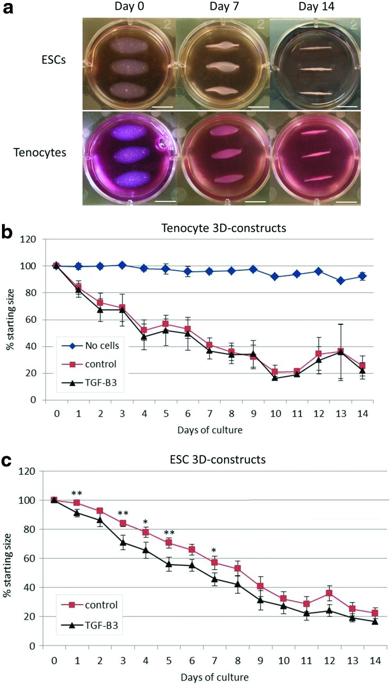
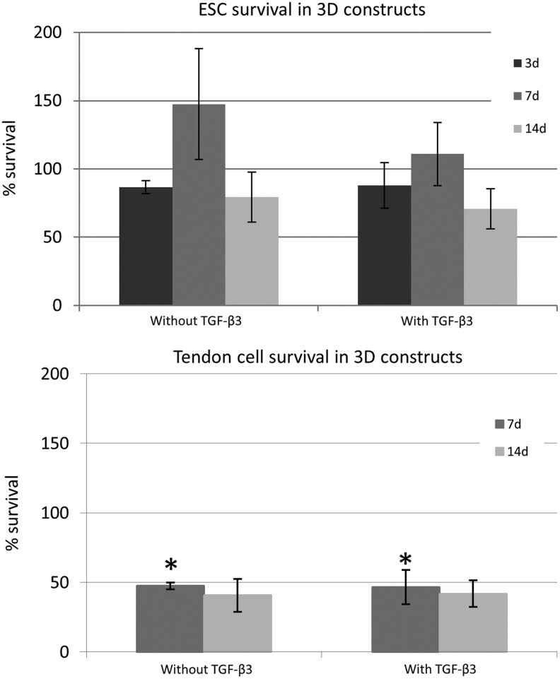
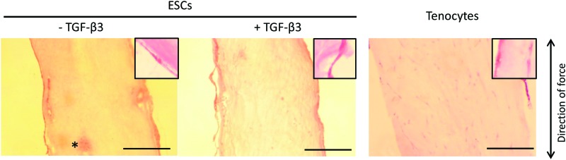
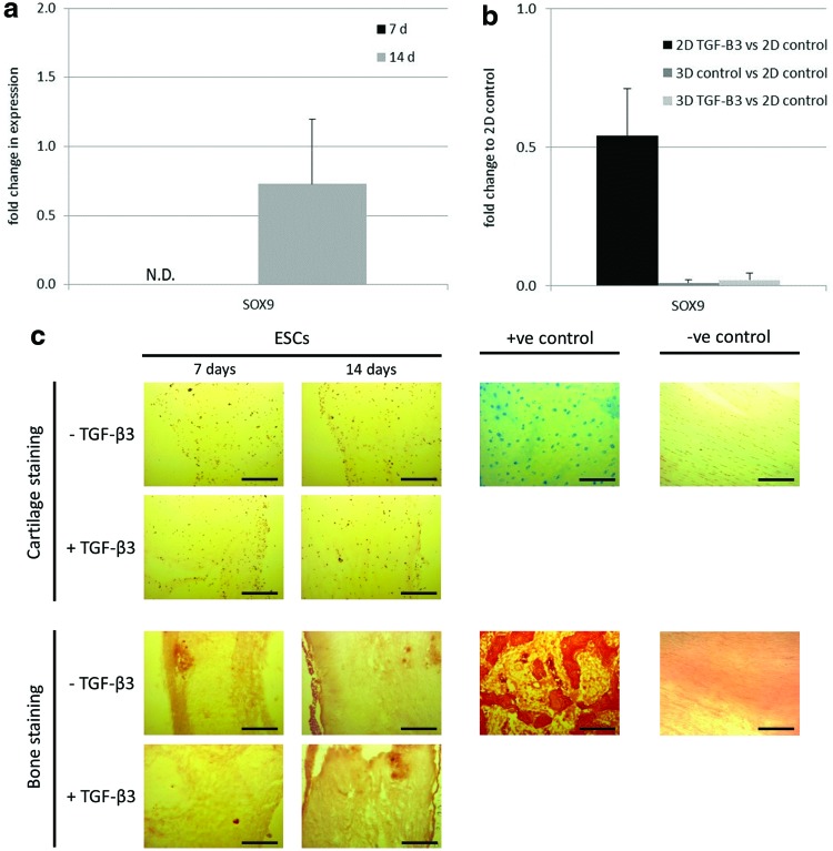
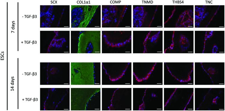
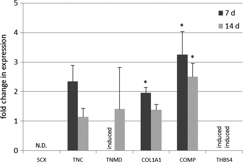
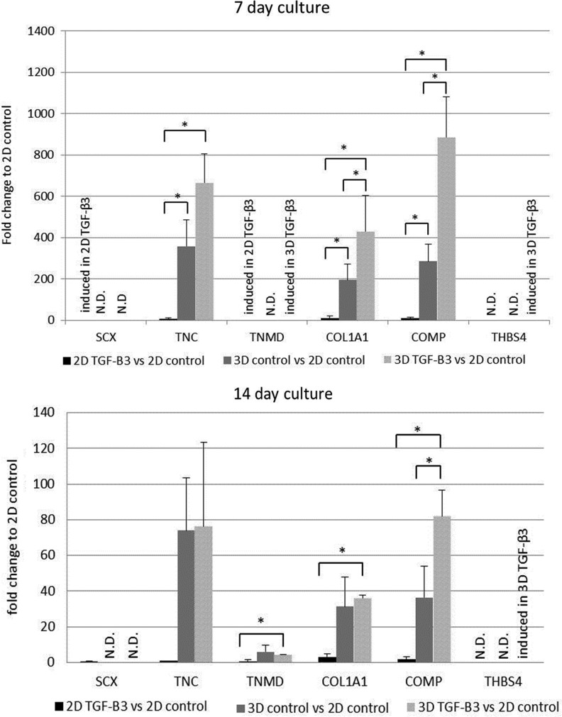
The natural reparative mechanisms triggered by tendon damage often lead to the formation of biomechanically inferior scar tissue that is prone to re-injury. Before the efficient application of stem cell-based regenerative therapies, the processes regulating tenocyte differentiation should first be better understood. Three-dimensional (3D) growth environments under strain and the exogenous addition of transforming growth factor beta3 (TGF-β3) have separately been shown to promote tendon differentiation. The aim of this study was to determine the ability of both of these factors to induce tendon differentiation of equine embryo-derived stem cells (ESCs). ESCs seeded into 3D collagen constructs can contract the matrix to a similar degree to that of tenocyte-seeded constructs and histologically appear nearly identical, with no areas of cartilage or bone tissue deposition. Tendon-associated genes and proteins Tenascin-C, Collagen Type I, and COMP are significantly up-regulated in the 3D ESC constructs compared with tenogenic induction in monolayer ESC cultures. The addition of TGF-β3 to the 3D cultures further up-regulates the expression of these genes and also induces the expression of mature tenocyte markers Tenomodulin and Thrombospondin-4. Our results show that when ESCs are exposed to the intrinsic forces exerted by a 3D culture environment, they express tendon-associated genes and proteins which are indicative of tenocyte lineage differentiation and that this effect is synergistically enhanced and accelerated by the addition of TGF-β3.


Figures

<b>FIG. 1.</b>
FIG. 1.
(a) Embryo-derived stem cell (ESC)-seeded constructs in three-dimensional (3D) culture contract the collagen matrix in a comparable manner to tenocyte-seeded constructs over the same time course. Scale bar=10 mm. (b) The contraction rates of tenocyte-seeded constructs are unaffected by the presence or absence of transforming growth factor beta3 (TGF-β3). (c) ESC-seeded constructs display enhanced rates of contraction in the presence of TGF-β3, which reach statistical significance over the first 5 days. **p<0.05, *p<0.1. Error bars represent the standard error of the mean. Color images available online at www.liebertpub.com/tea
<b>FIG. 2.</b>
FIG. 2.
Cell survival in ESC-seeded constructs is unaffected by the presence of TGF-β3 over the experimental time course. Cell survival of adult tendon cells is significantly lower than that of the ESCs after 7 days of culture *p<0.05. Error bars represent the standard error of the mean.
<b>FIG. 3.</b>
FIG. 3.
Hematoxylin and Eosin histological staining of sectioned 3D constructs show a high level of collagen fiber alignment after 14 days of culture. The majority of ES cells have migrated to the periphery of the construct and aligned with the direction of the anchoring tension. Inset boxes show magnified images of spindle cell morphology. No qualitative differences can be seen between ESC-constructs cultured in the presence or absence of TGF-β3. ESC-seeded constructs show a very high degree of similarity to tenocyte-seeded constructs cultured over the same period. *Remnants of initial ESC-seeded cluster. Scale bar=0.5 mm. Images shown are representative of three biological repeats. Color images available online at www.liebertpub.com/tea
<b>FIG. 4.</b>
FIG. 4.
(a) Quantitative PCR demonstrates that SOX9 expression is only detected in 14 day 3D ESC constructs, and its expression is not up-regulated in the presence of TGF-β3. N.D., expression not detected. (b) Relative to spontaneously differentiating two-dimensional (2D) ESC cultures, ESCs grown in the 3D environment (both with and without TGF-β3) for 14 days inhibit the expression of SOX9. Error bars represent the standard error of the mean. (c) Alcian Blue and Alizarin Red S staining, for cartilage and bone tissue components, respectively, show no positive staining in the ESC-seeded constructs over the 14 day time course, with or without TGF-β3 treatment. Positive controls were carried out with mature equine cartilage and bone sections. Negative controls used mature equine tendon sections. Scale bar=250 μm. Images shown are representative of three biological repeats. Color images available online at www.liebertpub.com/tea
<b>FIG. 5.</b>
FIG. 5.
Immunohistochemistry of longitudinal sections of 3D ESC-constructs shows the presence of tendon-associated proteins at both 7 and 14 days of culture. The intensity of each protein appears unaffected by the presence of TGF-β3, except for SCX, which shows stronger staining at 7 days under TGF-β3 treatment. DAPI staining of the nuclei is shown in blue. Data shown are representative of testing on three cell lines. Scale bar=40 μm. Images shown are representative of three biological repeats. Color images available online at www.liebertpub.com/tea
<b>FIG. 6.</b>
FIG. 6.
Quantitative PCR of 3D ESC constructs over 7 and 14 days demonstrates that the tendon-associated genes COL1α1 and COMP are significantly up-regulated at 7 days in the presence of TGF-β3 compared with untreated constructs. COMP expression remains significantly higher after 14 days. TNC also shows a trend for up-regulation in 3D ESC constructs in the presence of TGF-β3 after 7 days. The induction of THBS4 at 7 and 14 days, and the induction of TNMD at 7 days, only takes place in the TGF-β3-treated constructs. Induced=expression was only detected in the cultures in the presence of TGF-β and not in the control cultures at this time point. N.D., expression not detected. *p<0.05. Error bars represent the standard error of the mean.
<b>FIG. 7.</b>
FIG. 7.
Quantitative PCR analysis of tendon-associated genes in 3D ESC constructs (with and without TGF-β3) and 2D cultures in the presence of TGF-β3 displayed relative to 2D spontaneously differentiating ESC cultures. TNC, COL1α1, and COMP are significantly up-regulated in untreated 3D cultures compared with TGF-β3-treated 2D cultures. COL1α1 and COMP show further significant up-regulation when TGF-β3 is added to the 3D constructs, whereas TNC shows a trend for further up-regulation in 3D culture in the presence of TGF-β3 after 7 days. TNMD is only induced in the presence of TGF-β3 at 7 days, whereas THBS4 is only induced in 3D culture with TGF-β3 treatment. After 14 days, there is still a strong trend for increased tendon gene expression in 3D cultures compared with TGF-β3-treated 2D cultures, and the addition of TGF-β3 to 3D cultures results in a significant increase in the expression of TNMD, COL1α1, and COMP. THBS4 again is only expressed in 3D cultures in the presence of TGF-β3. Induced=expression was only detected in the cultures in the presence of TGF-β and not in the control cultures at this time point. N.D.=Expression not detected. *p<0.05. Error bars represent the standard error of the mean.

References

    1. Dyson S.J.Medical management of superficial digital flexor tendonitis: a comparative study in 219 horses (1992–2000). Equine vet J 36,415, 2004 - PubMed
    1. Cox D.A.Transforming growth factor-beta 3. Cell Biol Int 19,357, 1995 - PubMed
    1. Kuo C.K., Petersen B.C., and Tuan R.S.Spatiotemporal protein distribution of TGF-betas, their receptors, and extracellular matrix molecules during embryonic tendon development. Dev Dyn 237,1477, 2008 - PMC - PubMed
    1. Pryce B.A., et al. . Recruitment and maintenance of tendon progenitors by TGF beta signalling are essential for tendon formation. Development 136,1351, 2009 - PMC - PubMed
    1. Barsby T., and Guest D.Transforming growth factor beta3 promotes tendon differentiation of equine embryo-derived stem cells. Tissue Eng Part A 19,2156, 2013 - PubMed

Publication types

MeSH terms

Substances

LinkOut - more resources