. 2014 Mar-Jun;28(1-2):70-85.
doi: 10.3109/01677063.2013.878339. Epub 2014 Mar 17.

Molecular profiles of parvalbumin-immunoreactive neurons in the superior temporal cortex in schizophrenia

Affiliations

Molecular profiles of parvalbumin-immunoreactive neurons in the superior temporal cortex in schizophrenia

Charmaine Y Pietersen et al. J Neurogenet. 2014 Mar-Jun.

Abstract

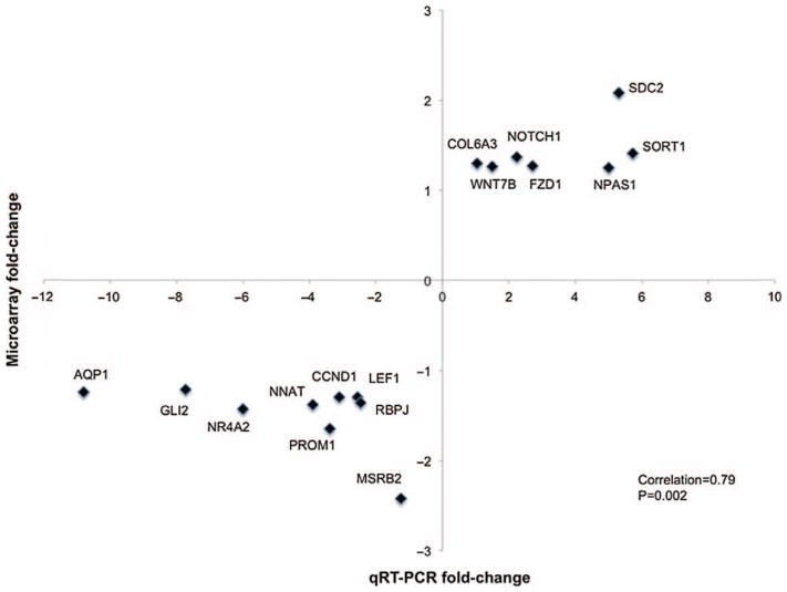
Dysregulation of pyramidal cell network function by the soma- and axon-targeting inhibitory neurons that contain the calcium-binding protein parvalbumin (PV) represents a core pathophysiological feature of schizophrenia. In order to gain insight into the molecular basis of their functional impairment, we used laser capture microdissection (LCM) to isolate PV-immunolabeled neurons from layer 3 of Brodmann's area 42 of the superior temporal gyrus (STG) from postmortem schizophrenia and normal control brains. We then extracted ribonucleic acid (RNA) from these neurons and determined their messenger RNA (mRNA) expression profile using the Affymetrix platform of microarray technology. Seven hundred thirty-nine mRNA transcripts were found to be differentially expressed in PV neurons in subjects with schizophrenia, including genes associated with WNT (wingless-type), NOTCH, and PGE2 (prostaglandin E2) signaling, in addition to genes that regulate cell cycle and apoptosis. Of these 739 genes, only 89 (12%) were also differentially expressed in pyramidal neurons, as described in the accompanying paper, suggesting that the molecular pathophysiology of schizophrenia appears to be predominantly neuronal type specific. In addition, we identified 15 microRNAs (miRNAs) that were differentially expressed in schizophrenia; enrichment analysis of the predicted targets of these miRNAs included the signaling pathways found by microarray to be dysregulated in schizophrenia. Taken together, findings of this study provide a neurobiological framework within which hypotheses of the molecular mechanisms that underlie the dysfunction of PV neurons in schizophrenia can be generated and experimentally explored and, as such, may ultimately inform the conceptualization of rational targeted molecular intervention for this debilitating disorder.

Keywords: cerebral cortex; gene expression profiling; laser capture microdissection; microRNA.


Conflict of interest statement

Declaration of interest: The authors report no conflicts of interest. The authors alone are responsible for the content and writing of the paper.

Figures

Figure 1
Figure 1
Identification of PV neurons, representative virtual gels of amplified RNA products, and heatmap of 739 differentially expressed genes. (A) Photomicrograph of PV-immunoreactive neurons. Scale bar = 10 μm. (B) Representative virtual gels showing the sizes of products after two rounds of linear amplification of RNA extracted from PV-immunoreactive neurons from a normal control (C) and a schizophrenia (S) subjects. (C) Heatmap of the 739 differentially expressed genes in schizophrenia compared with normal control subjects, showing the segregation of genes based on diagnosis under the stringency criteria of fold-change >1.2, FDR-adjusted p<0.05.
Figure 2
Figure 2
Correlation analysis comparing fold-changes of selected genes determined by microarray and qRT-PCR. Comparison of fold-changes of randomly selected genes (N = 10) and genes selected from the most significantly affected pathways (N = 6) determined by microarray and qRT-PCR. AQP1, aquaporin 1; CCND1, cyclin D1; COL6A3, collagen, type VI, alpha 3; FZD1, frizzled family receptor 1; GLI2, glioma-associated oncogene family zinc finger 2; LEF1, lymphoid enhancer-binding factor 1; MSRB2, methionine sulfoxide reductase B2; NNAT, neuronatin; NOTCH1, notch1; NPAS1, neuronal PAS domain protein 1; NR4A2, nuclear receptor subfamily 4, group A, member 2; PROM1, prominin 1; RBPJ, recombinant signal binding protein for immunoglobulin kappa J region; SDC2, syndecan 2; SORT1, sortilin; WNT7B, wingless-type MMTV integration site family, member 7B.
Figure 3
Figure 3
Schematic diagram showing the convergence of WNT and NOTCH signaling onto cell cycle regulation. Modified GeneGo pathway diagram showing that aberrant WNT and NOTCH signaling in schizophrenia may contribute to cell cycle dysregulation (see text for details). FZD1, frizzled family receptor 1; EGFR, epidermal growth factor receptor; LEF1, lymphoid enhancer-binding factor 1; TCF, transcription factor 1; RBPJ, recombination signal binding protein for immunoglobulin kappa J region.

References

    1. Ables JL, Breunig JJ, Eisch AJ, Rakic P. Not(ch) just development: Notch signalling in the adult brain. Nat Rev Neurosci. 2011;12:269–283. - PMC - PubMed
    1. Ahmad-Annuar A, Ciani L, Simeonidis I, Herreros J, Fredj NB, Rosso SB, et al. Signaling across the synapse: A role for Wnt and Dishevelled in presynaptic assembly and neurotransmitter release. J Cell Biol. 2006;174:127–139. - PMC - PubMed
    1. Anderson MF, Aberg MA, Nilsson M, Eriksson PS. Insulin-like growth factor-I and neurogenesis in the adult mammalian brain. Brain Res Dev Brain Res. 2002;134:115–122. - PubMed
    1. Behrens MM, Sejnowski TJ. Does schizophrenia arise from oxidative dysregulation of parvalbumin-interneurons in the developing cortex? Neuropharmacology. 2009;57:193–200. - PMC - PubMed
    1. Benes FM. Regulation of cell cycle and DNA repair in post-mitotic GABA neurons in psychotic disorders. Neuropharmacology. 2011;60:1232–1242. - PubMed

Publication types

MeSH terms

LinkOut - more resources