Review
. 2014 Mar 12;15(3):266-82.
doi: 10.1016/j.chom.2014.02.011.

Human viral oncogenesis: a cancer hallmarks analysis

Affiliations
Review

Human viral oncogenesis: a cancer hallmarks analysis

Enrique A Mesri et al. Cell Host Microbe. .

Abstract

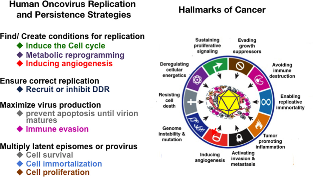
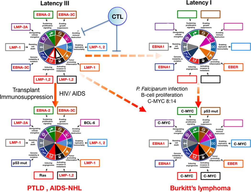
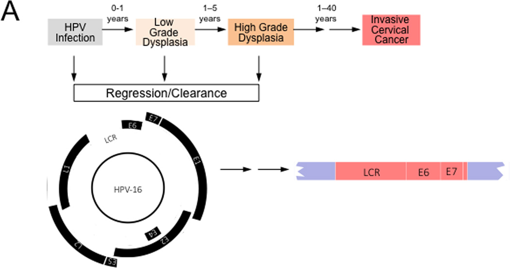
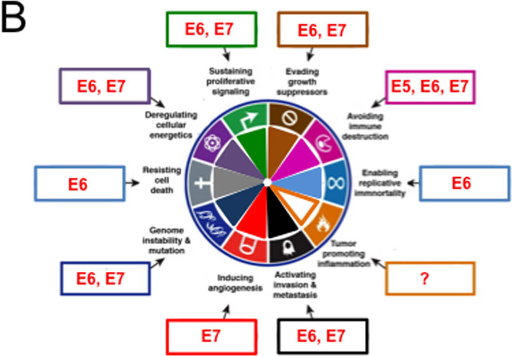
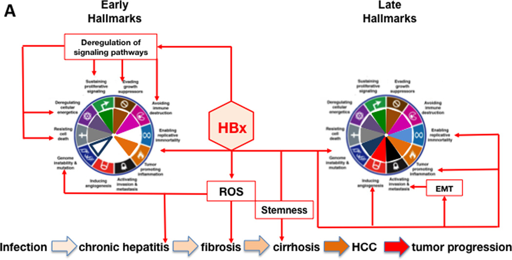
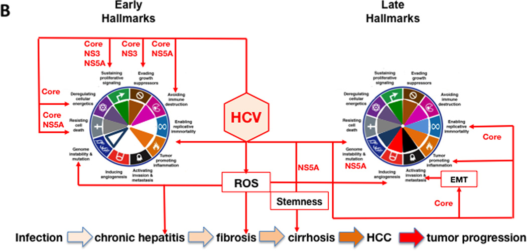
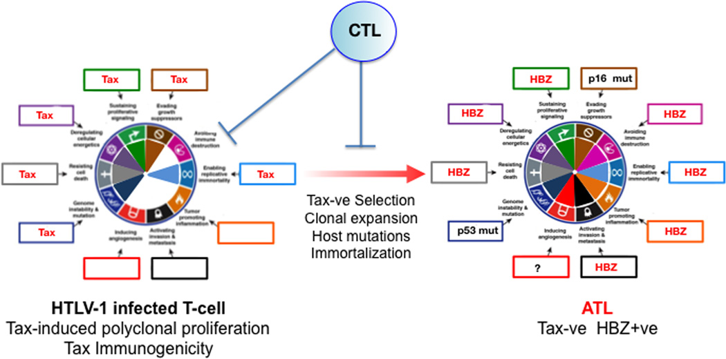
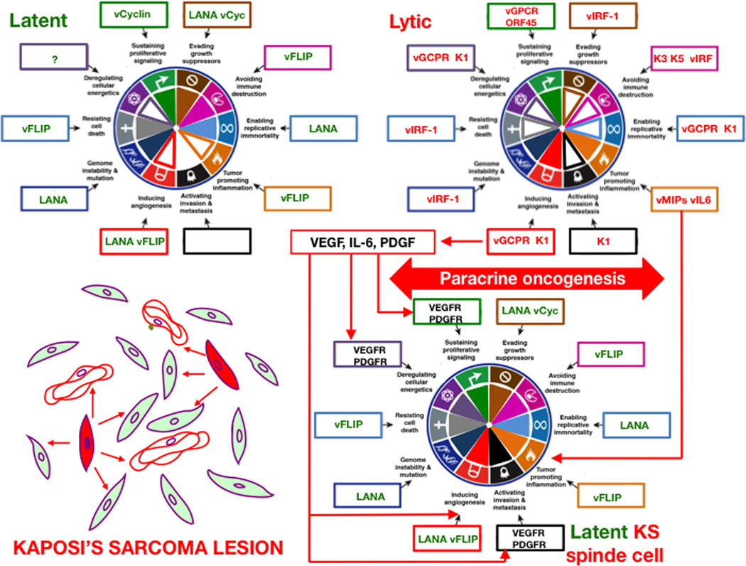
Approximately 12% of all human cancers are caused by oncoviruses. Human viral oncogenesis is complex, and only a small percentage of the infected individuals develop cancer, often many years to decades after the initial infection. This reflects the multistep nature of viral oncogenesis, host genetic variability, and the fact that viruses contribute to only a portion of the oncogenic events. In this review, the Hallmarks of Cancer framework of Hanahan and Weinberg (2000 and 2011) is used to dissect the viral, host, and environmental cofactors that contribute to the biology of multistep oncogenesis mediated by established human oncoviruses. The viruses discussed include Epstein-Barr virus (EBV), high-risk human papillomaviruses (HPVs), hepatitis B and C viruses (HBV and HCV, respectively), human T cell lymphotropic virus-1 (HTLV-1), and Kaposi's sarcoma herpesvirus (KSHV).


Figures

Figure 1
Figure 1. Oncogenic risks of viral replication and persistence strategies
Many of the molecular mechanisms deployed by human oncoviruses to maximize replication and persistence imply hijacking the host cell’s signaling machinery leading to acquisition of cancer hallmarks. The main strategies for viral replication and persistence are listed (on the left). Below each of them are the cellular responses that the virus induces to undertake each of these strategies. Virally induced cell responses are color coded according to the Hallmarks of Cancer to which they correspond (on the right). The picture on the right is reproduced from the review from Hanahan and Weinberg (2011).
Figure 2
Figure 2. Hallmarks of Cancer analysis of EBV mediated lymphomagenesis
When EBV infects B-cells it establishes a “growth program” or Latency III pattern, which expresses EBV proteins with sufficient oncogenic potential to cause immortalization in vitro, and B-cell growth and differentiation in vivo. However this program is too immunogenic due to EBNA-2,3 protein expression. The resulting immune pressure from cytotoxic T lymphocytes (CTL) promotes virus first switching to the default program (see text) and then the latency I programs that is not immunogenic but it is also not oncogenic since it only expresses EBNA1 and EBER. In immunosuppressed and AIDS patients Lat III cells can thrive and lead to PTLD or AIDS-NHL where host somatic mutations such as Bcl-6 overexpression, p53 mutation or Ras mutations further contributes to all the hallmarks for lymphomagenesis. In malaria affected regions P. falciparum induced B cell proliferation favors the occurrence of 8:14 translocations that lead to c-myc overexpression. c-myc expression with further host somatic mutations such as p53 inactivation may complement the oncogenicity of Latency I pattern leading to Burkitt’s lymphomagenesis. Viral genes are depicted in red and host genes in black. The figure is not a strict representation of experimental data but rather a compilation of published information analyzed in the context of the Hallmarks of Cancer. Genes chosen for hallmarks activation represent one of the available examples and are based on published data with preference for tumor relevant systems where available. Filled portions within the hallmark pie are used to represent stronger or well-documented hallmark activation. Empty portions represent weaker effects or lack of activation/evidence. See text for explanations and references.
Figure 3
Figure 3. Hallmarks of Cancer analysis of HPV associated cervical carcinogenesis
High-risk HPV infections can give rise to low-grade dysplasia (also referred to as cervical intraepithelial neoplasia 1 (CIN1)), which can progress to high-grade dysplasia (also referred to as CIN2/3). Many of these lesions spontaneously regress, presumably because of immune clearance by the host. These lesions contain episomal HPV genomes and expression of the viral genes is tightly controlled by the interplay of cellular and viral factors. Malignant progression to invasive cervical cancer is often a very slow process and cervical cancers can arise years or decades after the initial infection. Cervical carcinomas frequently contain integrated HPV sequences. Expression of the viral E2 transcriptional repressor is generally lost upon viral genome integration resulting in dysregulated viral gene expression from the viral Long Control Region (LCR). HPV E6 and E7 are consistently expressed even after genome integration and expression of these proteins is necessary for the maintenance of the transformed phenotype. See text for detail. (B) High-risk HPV proteins target almost the entire spectrum of the Cancer Hallmarks. HPV genes that affect each hallmark are indicated within boxes around the wheel. See text for details
Figure 4
Figure 4. Hallmarks of Cancer activation during the progression of HBV and HCV infection to HCC
The diagram shows hallmarks activation during HBV infection, primarily by the viral HBx protein (panel A), and by HCV infection, mainly by the viral core, NS5A and NS3 proteins (panel B). The host mediators that contribute to each of the hallmarks are indicated in black text boxes. Hallmarks achieved early (left wheel) and late (right wheel) over the course of HBV- and HCV-associated HCC pathogenesis is shown for each panel. The major steps in the progression of HBV and HCV infection to HCC are shown at the bottom of each panel. The progression of these steps is indicated by increasingly darker tan-colored arrows. Please see the text for additional details.
Figure 5
Figure 5. Hallmarks of Cancer analysis for HTLV-1 induced Adult T-cell Lymphoma
Tax activation of survival and proliferation hallmarks in HTLV-1 infected T-cells leads to polyclonal expansion of infected cells leading to immortalization. However since Tax is immunogenic, host CTLs select for infected cells with downregulated or deleted Tax. After a prolonged asymptomatic period of 20–40 years, that involves acquisition of host mutations, immortalization and clonal expansion, adult T-cell leukemia (ATL) cells emerge in approximately 5% of infected individuals. Tax functions are compensated by HZB-mediated hallmark activation and by host mutations such as p16INK4A and p53. Most HTLV-1-infected individuals remain asymptomatic carriers.
Figure 6
Figure 6. Hallmarks of Cancer Analysis for Kaposi’s sarcoma pathogenesis based on the paracrine oncogenesis hypothesis
KSHV infections could be either latent or lytic. Latently infected cells express the LANA transcript and the KSHV miRNA. Although these genes can activate KS cancer hallmarks they are insufficient to transform the cell and to induce the characteristic KS angiogenic phenotype. Lytic infected cells on the other hand express viral genes that make the highly angiogenic and inflammatory but they are doomed for cytopathic death and under immune control. In immunosuppressed and AIDS individuals, lytic infection is more permissible and it can combine with latently infected cells to form tumors via the mechanism of paracrine oncogenesis (see text for details). Bottom left is a schematic representation of a KS tumor showing latently infected cells (filled in green), lytically infected cells (filled in red), blood vessels (unfilled in red) and paracrine effects (depicted as red arrows). Within the boxes around the wheel, latent viral genes are in green, lytic viral genes in red and host genes are in black. The figure is not a strict representation of experimental data but a compilation of published information analyzed in the context of the Hallmarks of Cancer and the paracrine oncogenesis hypothesis. Genes chosen for hallmarks activation represent one of the available examples and are based on published data with preference for tumor relevant systems where available. Filled portions of hallmark pie are used to represent stronger or well documented hallmark activation. Empty portions represent weaker effects or lack of activation/ evidence. See text for explanations and references.

References

    1. Aoki Y, Jaffe ES, Chang Y, Jones K, Teruya-Feldstein J, Moore PS, Tosato G. Angiogenesis and hematopoiesis induced by Kaposi's sarcoma-associated herpesvirus-encoded interleukin-6. Blood. 1999;93:4034–4043. - PubMed
    1. Aoki Y, Tosato G. Interactions between HIV-1 Tat and KSHV. Current topics in microbiology and immunology. 2007;312:309–326. - PubMed
    1. Arvey A, Tempera I, Tsai K, Chen HS, Tikhmyanova N, Klichinsky M, Leslie C, Lieberman PM. An atlas of the Epstein-Barr virus transcriptome and epigenome reveals host-virus regulatory interactions. Cell host & microbe. 2012;12:233–245. - PMC - PubMed
    1. Arzumanyan A, Friedman T, Ng IO, Clayton MM, Lian Z, Feitelson MA. Does the hepatitis B antigen HBx promote the appearance of liver cancer stem cells? Cancer research. 2011;71:3701–3708. - PMC - PubMed
    1. Arzumanyan A, Reis HM, Feitelson MA. Pathogenic mechanisms in HBV- and HCV-associated hepatocellular carcinoma. Nature reviews Cancer. 2013;13:123–135. - PubMed

Publication types