Review
. 2014 Mar 12;15(3):295-305.
doi: 10.1016/j.chom.2014.02.003.

Host immune response to infection and cancer: unexpected commonalities

Affiliations
Review

Host immune response to infection and cancer: unexpected commonalities

Romina S Goldszmid et al. Cell Host Microbe. .

Abstract

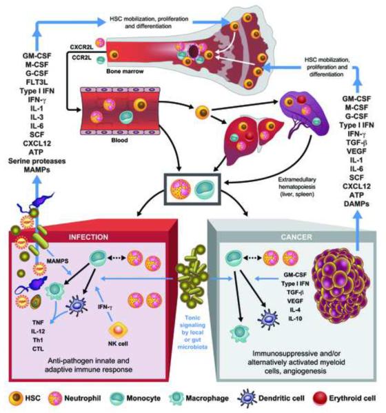
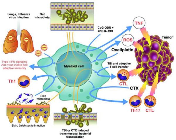
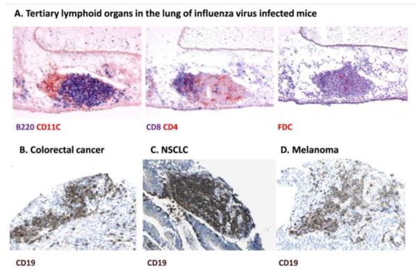
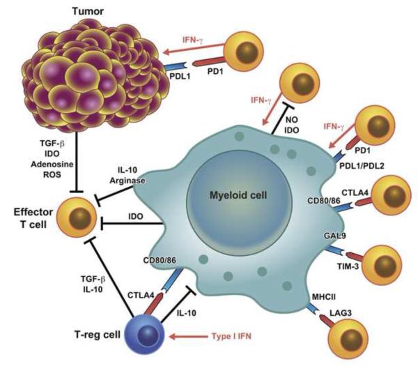
Both microbes and tumors activate innate resistance, tissue repair, and adaptive immunity. Unlike acute infection, tumor growth is initially unapparent; however, inflammation and immunity affect all phases of tumor growth from initiation to progression and dissemination. Here, we discuss the shared features involved in the immune response to infection and cancer including modulation by commensal microbiota, reactive hematopoiesis, chronic immune responses and regulatory mechanisms to prevent collateral tissue damage. This comparative analysis of immunity to infection and cancer furthers our understanding of the basic mechanisms underlying innate resistance and adaptive immunity and their translational application to the design of new therapeutic approaches.


Figures

Figure 1
Figure 1
Reactive hematopoiesis in infection and cancer. Factors produced by infected tissues or tumors and their stroma induce mobilization of HSCs from endosteal/arteriolar niches to vascular niches and promote their proliferation and differentiation. In response to chemotactic stimuli, HSCs, monocytes and neutrophils marginalize in the blood circulation. HSCs reach organs such as liver and spleen where extramedullary hematopoiesis occurs. Neutrophils and inflammatory monocytes enter infected tissues and tumor stroma, where monocytes differentiate into macrophages- or DC-like cells. In infections, these inflammatory myeloid cell types, in part in response to MAMPs and to IFN-γ secreted by NK cells, produce cytokines that favor the generation of effector Th cells, often Th1, and CTL. In tumors various mediators present in the microenvironment lead to the appearance of myeloid cells with immunosuppressive activity and favoring tumor growth and angiogenesis. In both infections and cancer, the differentiation of the myeloid cells in the inflamed tissue is modulated by the presence of the gut microbiota. Myeloid cells with immunosuppressive activities are also observed in infections where they may contribute to resolution of inflammation and immunity, while myeloid cells in the tumor can become immunostimulating if properly stimulated and the activity of suppressive factors such as IL-10 is inhibited.
Figure 2
Figure 2
Contribution of the commensal microbiota to the resistance to infection and anti-cancer therapy effectiveness. The presence of the gut microbiota is essential for resistance in the lungs to respiratory virus infection and for the success of anti-cancer therapies in subcutaneous tumors. The microbiota controls the efficacy of CpG-oligonucleotide (ODN) therapy combined with anti-IL-10R antibody treatment by enabling myeloid cells to produce TNF and the early genotoxic effect of oxaliplatin by enabling them to release ROS. Gut transmucosal translocation of bacteria induced by total body irradiation (TBI) or cyclophosphamide (CTX) treatment is required for the anti-cancer effectiveness of adoptive T cell transfer and for the induction of adaptive immunity following CTX-induced immunogenic cell death. The skin microbiota control skin immunity in a compartmentalized way and it is required for an efficient immune response against L. major infection.
Figure 3
Figure 3
Tertiary lymphoid organs (TLOs) in infection and cancer. A. In chronic infections such as in the lung of influenza virus infected mice shown here, TLOs are present and characterized by anatomically contiguous but separated T (CD4 or CD8 positive) and B cell areas (B220 positive) and the presence of conventional (CD11C positive) or follicular (FDC) DCs, suggesting localized sites of T and B cell responses (Reproduced with permission from ©2009 GeurtsvanKessel et al. Originally published in Journal of Experimental Medicine: 206:2339-2349. doi: 10.1084/jem.20090410). C-D. TLOs are often present in tumors as visualized by the B cell marker CD19 (from www.proteinatlas.org).
Figure 4
Figure 4
Main immune checkpoints negatively regulating the immune response in infection and cancer. T cells in the tumor environment have an exhausted phenotype and their proliferation and activation is restrained both by tissue intrinsic tolerogenic mechanisms that limit excessive inflammatory and immune response to infection as well as by the mechanisms responsible for the contraction of the immune response to infection.

References

    1. Abt MC, Osborne LC, Monticelli LA, Doering TA, Alenghat T, Sonnenberg GF, Paley MA, Antenus M, Williams KL, Erikson J, et al. Commensal bacteria calibrate the activation threshold of innate antiviral immunity. Immunity. 2012;37:158–170. - PMC - PubMed
    1. Anderson AC. Tim-3, a negative regulator of anti-tumor immunity. Curr Opin Immunol. 2012;24:213–216. - PubMed
    1. Ascensao JL, Oken MM, Ewing SL, Goldberg RJ, Kaplan ME. Leukocytosis and large cell lung cancer. A frequent association. Cancer. 1987;60:903–905. - PubMed
    1. Baldridge MT, King KY, Goodell MA. Inflammatory signals regulate hematopoietic stem cells. Trends Immunol. 2011;32:57–65. - PMC - PubMed
    1. Barber DL, Wherry EJ, Masopust D, Zhu B, Allison JP, Sharpe AH, Freeman GJ, Ahmed R. Restoring function in exhausted CD8 T cells during chronic viral infection. Nature. 2006;439:682–687. - PubMed