. 2014 Apr;184(4):1230-1239.
doi: 10.1016/j.ajpath.2013.12.023. Epub 2014 Mar 12.

VEGFA activates erythropoietin receptor and enhances VEGFR2-mediated pathological angiogenesis

Affiliations

VEGFA activates erythropoietin receptor and enhances VEGFR2-mediated pathological angiogenesis

Zhihong Yang et al. Am J Pathol. 2014 Apr.

Abstract

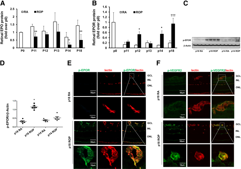
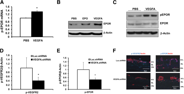
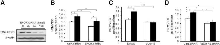
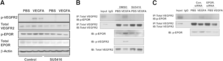
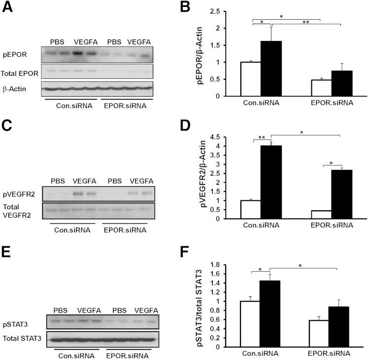
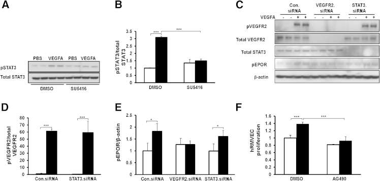
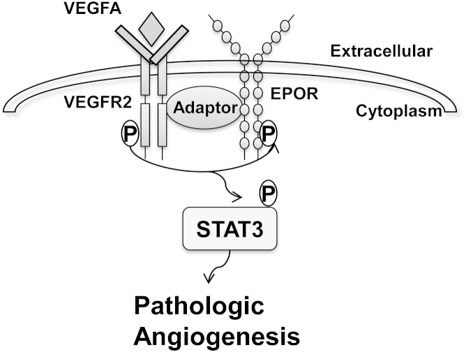
Clinical and animal studies implicate erythropoietin (EPO) and EPO receptor (EPOR) signaling in angiogenesis. In the eye, EPO is involved in both physiological and pathological angiogenesis in the retina. We hypothesized that EPOR signaling is important in pathological angiogenesis and tested this hypothesis using a rat model of oxygen-induced retinopathy that is representative of human retinopathy of prematurity. We first determined that EPOR expression and activation were increased and that activated EPOR was localized to retinal vascular endothelial cells (ECs) in retinas at postnatal day 18 (p18), when pathological angiogenesis in the form of intravitreal neovascularization occurred. In human retinal microvascular ECs, EPOR was up-regulated and activated by VEGF. Lentiviral-delivered shRNAs that knocked down Müller cell-expressed VEGF in the retinopathy of prematurity model also reduced phosphorylated EPOR (p-EPOR) and VEGFR2 (p-VEGFR2) in retinal ECs. In human retinal microvascular ECs, VEGFR2-activated EPOR caused an interaction between p-EPOR and p-VEGFR2; knockdown of EPOR by siRNA transfection reduced VEGF-induced EC proliferation in association with reduced p-VEGFR2 and p-STAT3; however, inhibition of VEGFR2 activation by siRNA transfection or semaxanib (SU5416) abolished VEGFA-induced proliferation of ECs and phosphorylation of VEGFR2, EPOR, and STAT3. Our results show that VEGFA-induced p-VEGFR2 activates EPOR and causes an interaction between p-EPOR and p-VEGFR2 to enhance VEGFA-induced EC proliferation by exacerbating STAT3 activation, leading to pathological angiogenesis.


Figures

Figure 1
Figure 1
EPOR expression and activation increased in vascular endothelium in rat ROP model. A and B: Protein analyses for EPO (A) and EPOR (B) in retinas from rat pups raised in room air (RA) and ROP model pups at time points from baseline to p18. C and D: Representative Western blots of p-EPOR in retina at p14 and p18 (C) and quantification of gels (D). E and F: Immunohistochemical staining of p-EPOR (green) (E) or p-VEGFR2 (green) (F) colabeling with lectin (red) in retinal cryosections at p18. The boxed region in each upper row corresponds to the adjacent image at higher magnification in the lower row. Negative controls are shown in Supplemental Figure S1A. Data are expressed as means ± SD (A and B) or as individual data points with means ± SD (D). P < 0.05, ∗∗P < 0.01 versus RA at the same developmental age. †††P < 0.001, overall analysis of variance. Scale bars: 50 μm (E and F, upper rows); 10 μm (E and F, lower rows). GCL, ganglion cell layer; INL, inner nuclear layer; ONL, outer nuclear layer.
Figure 2
Figure 2
EPOR is up-regulated and activated by VEGFA in vitro and in vivo. A and B: qPCR of human EPOR (A) and Western blots of EPOR protein in hRMVECs stimulated by 20 ng/mL VEGFA or 10 IU/mL EPO for 18 hours (B). Human β-actin was used as an internal control. C: Western blots of p-EPOR and total EPOR in hRMVECs stimulated with 20 ng/mL VEGFA or PBS for 30 minutes. D and E: Quantification of Western blots of p-VEGFR2 (D) and p-EPOR (E) in retina. F: IHC of p-VEGFR2 or p-EPOR (blue) colabeled with lectin (red) in retinal sections from rat pups injected with lentivirus-delivered luciferase shRNA (Luc.shRNA) or VEGFA shRNA (VEGFA.shRNA) at p18 in rat ROP model. Negative controls are shown in Supplemental Figure S1B. Data expressed as means ± SD, representative of three or more independent experiments with n = 2-3 for each experiment. P < 0.05 versus control. Scale bar = 50 μm.
Figure 3
Figure 3
Knockdown of EPOR reduces VEGFA-induced EC proliferation, and inhibition of VEGFR2 activation inhibits it, in hRMVECs. A: Western blots of total EPOR in hRMVECs transfected with 0 to 100 pmol EPOR siRNA. B–D: VEGFA-induced proliferation assay in hRMVECs transfected with control siRNA (Con.siRNA) or EPOR siRNA (B), in hRMVECs treated with SU5416 (C), and in hRMVECs transfected with Con.siRNA or VEGFR2 siRNA (D). Data are expressed as means ± SD, representative of three or more independent experiments. White bars, PBS; black bars, VEGFA (B–D). P < 0.05, ∗∗∗P < 0.001.
Figure 4
Figure 4
VEGFA-activated EPOR interacts with p-VEGFR2 in hRMVECs. A: Western blots of phosphorylated and total VEGFR2 and EPOR in hRMVECs treated with PBS or VEGFA in the presence of DMSO control or SU5416. B and C: Coimmunoprecipitation of p-EPOR and VEGFR2 in hRMVECs treated with PBS or VEGFA in the presence of DMSO control or SU5416 (B) or in hRMVECs transfected with Con.siRNA or EPOR.siRNA (C). Gels are representative of three or more independent experiments with n = 2 for each experiment. IB, immunoblot (Western blot); IP, immunoprecipitation.
Figure 5
Figure 5
Knockdown of EPOR reduces VEGFA-induced p-VEGFR2 and inhibits VEGFA-induced p-EPOR and p-STAT3 in hRMVECs. Representative Western blots, with quantification of densitometry, for p-EPOR (A and B), p-VEGFR2 (C and D), and p-STAT3 (E and F) in hRMVECs transfected with Con.siRNA or EPOR.siRNA and treated with PBS or VEGFA. Data are expressed as means ± SD, representative of three or more independent experiments with n = 2 for each experiment. White bars, PBS; black bars, VEGFA (in panels B, D, and F). P < 0.05, ∗∗P < 0.01.
Figure 6
Figure 6
STAT3 is the downstream target of activated VEGFR2 and EPOR, and activation of STAT3 mediates VEGFA-induced EC proliferation. A and B: Representative Western blots (A) and quantification (B) of p-STAT3 and total STAT3 in hRMVECs treated with PBS or VEGFA in the presence of control DMSO or SU5416. Quantification of densitometry of p-STAT3 is normalized to total STAT3. C–E: Representative Western blots (C) with quantification (D and E) of p-VEGFR2, p-EPOR, total VEGFR2, and total STAT3 in hRMVECs transfected with Con.siRNA, VEGFR2.siRNA, or STAT3.siRNA and treated with PBS or with 20 ng/mL VEGFA for 30 minutes. Quantification of densitometry of p-VEGFR2 is normalized to total VEGFR2 (D) and that of p-EPOR is normalized to β-actin (E). F: Cell proliferation assay in hRMVECs treated with PBS or VEGFA in the presence of DMSO or AG490 Data are expressed as means ± SD, representative of three or more independent experiments with n = 2 for each experiment. White bars, PBS; black bars, VEGFA (in panels B, D, E and F). P < 0.05, ∗∗∗P < 0.001.
Figure 7
Figure 7
The hypothetical signaling pathway in pathological angiogenesis regulated by interactions of activated VEGFR2 and EPOR. VEGFA activates VEGFR2, which then phosphorylates EPOR and forms an interaction with p-EPOR to exacerbate STAT3 activation and mediate pathological angiogenesis as seen in phase II ROP.

References

    1. Gilbert C. Retinopathy of prematurity: a global perspective of the epidemics, population of babies at risk and implications for control. Early Hum Dev. 2008;84:77–82. - PubMed
    1. Hartnett M.E., Penn J.S. Mechanisms and management of retinopathy of prematurity. N Engl J Med. 2012;367:2515–2526. - PMC - PubMed
    1. Smith L.E.H., Wesolowski E., McLellan A., Kostyk S.K., D’Amato R., Sullivan R., D’Amore P.A. Oxygen induced retinopathy in the mouse. Invest Ophthalmol Vis Sci. 1994;35:101–111. - PubMed
    1. Ashton N. Retrolental fibroplasia now retinopathy of prematurity. Br J Ophthalmol. 1984;68:689.
    1. Shah P.K., Narendran V., Kalpana N. Aggressive posterior retinopathy of prematurity in large preterm babies in South India. Arch Dis Child Fetal Neonatal Ed. 2012;97:F371–F375. - PubMed

Publication types

MeSH terms

Substances