. 2014 Mar 13;156(6):1235-1246.
doi: 10.1016/j.cell.2014.01.056.

S-glutathionylation of cryptic cysteines enhances titin elasticity by blocking protein folding

Affiliations

S-glutathionylation of cryptic cysteines enhances titin elasticity by blocking protein folding

Jorge Alegre-Cebollada et al. Cell. .

Abstract

The giant elastic protein titin is a determinant factor in how much blood fills the left ventricle during diastole and thus in the etiology of heart disease. Titin has been identified as a target of S-glutathionylation, an end product of the nitric-oxide-signaling cascade that increases cardiac muscle elasticity. However, it is unknown how S-glutathionylation may regulate the elasticity of titin and cardiac tissue. Here, we show that mechanical unfolding of titin immunoglobulin (Ig) domains exposes buried cysteine residues, which then can be S-glutathionylated. S-glutathionylation of cryptic cysteines greatly decreases the mechanical stability of the parent Ig domain as well as its ability to fold. Both effects favor a more extensible state of titin. Furthermore, we demonstrate that S-glutathionylation of cryptic cysteines in titin mediates mechanochemical modulation of the elasticity of human cardiomyocytes. We propose that posttranslational modification of cryptic residues is a general mechanism to regulate tissue elasticity.


Figures

Figure 1
Figure 1. Ig domains in the I-band of titin are rich in cryptic cysteines
(A) Schematic representation of a half-sarcomere. Titin (in color) extends from the Z-disk to the M-band of the sarcomere. The approximate location of the I91 domain is indicated with an asterisk. (B) Average cysteine content in the titin sequence. (C) Structure of the titin I91 domain (also known as I27, PDB code 1tit), highlighting in red its two cysteine residues. (D) The cysteines in I91 are cryptic and can only be S-glutathionylated after unfolding. A SUMO-I91 construct was incubated with GSSG for 1 h at different temperatures. S-glutathionylation was determined by western blotting and labeling with anti-glutathione antibodies (α-GSH). Ponceau S shows the total protein content. See also Figure S1
Figure 2
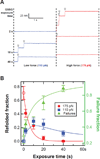
Figure 2. S-glutathionylation of cryptic cysteines inhibits refolding of I91
(A) I91 contains two cryptic cysteines that become exposed by mechanical unfolding. At a force of 130–175 pN, the unfolding of an I91 domain can be identified as a 25 nm step increase in end-to-end length. Exposure of the unfolded protein to GSSG leads to S-glutathionylation. (B) A single polyprotein is unfolded at 175 pN in 100 mM GSSG and then held extended for a variable length of time (exposure time; 2 s top trace, 40 s bottom trace). The force is then quenched to zero for 5 s to allow the protein to refold. We measure the extent of refolding by applying a probe pulse. (C) Refolded fraction as a function of exposure time for I918 polyproteins exposed to buffer only (downward triangles, N>35), 100 mM GSSG (solid circles, N>80), 200 mM GSH (upward triangles, N>80); and refolded fraction in the presence of 100 mM GSSG for a mutant I918 polyprotein where all cysteine residues were replaced by alanines (ΔCys, squares, N>120). The refolded fraction was calculated as the ratio between the number of unfolding steps detected during the probe pulse and the number of unfolding steps detected during the exposure pulse. Open symbols represent total refolding of I91 (weak + strong domains) in the presence of 100 mM GSSG, as determined using the double probe pulse protocol in Fig 3. Error bars represent SEM.
Figure 3
Figure 3. Graded mechanical modulation of I91 by S-glutathionylation
(A) Experimental recordings showing unfolding steps during the probe pulse (open arrowheads). Each recording was obtained in 100 mM GSSG after a different exposure time followed by 5 s refolding. Refolded domains with different mechanical stability were detected using a two-level probe pulse (110 pN in blue followed by 175 pN in red). (B) Refolded fraction (circles and squares, left axis) and failures (triangles, right axis) as a function of exposure time (N>40). Squares indicate weak domains detected during the low force regime; circles indicate native domains detected in the high force regime. Data points at zero exposure time were obtained in buffer without GSSG (open symbols, N=24). Solid lines were obtained from the kinetic model shown in Figure 5A. Error bars represent SEM. See also Figure S2
Figure 4
Figure 4. S-glutathionylation of different cysteines produces distinct mechanical phenotypes
(A) The single-cysteine mutant I91-Cys63 was mechanically unfolded and exposed to 100 mM GSSG for a variable amount of time. After a 5 s quench to allow for folding, refolding was probed using a linear force ramp up to 240 pN. (B) Mechanical stability of refolded I91-Cys63 after varying exposure times to 100 mM GSSG (N>25). Solid lines indicate fits of Gaussians with fixed widths and centroids. (C) Refolded fraction of I91-Cys47 (triangles, N>120) and I91-Cys63 (weak domains, squares; strong domains, circles; N>150) at different exposure times for a fixed quench time of 5 s in 100 mM GSSG. Data points at zero exposure time were obtained in the absence of GSSG (open symbols; N>150). Reaction rates of S-glutathionylation were measured from exponential fits (solid lines). Error bars represent SEM. See also Figure S3.
Figure 5
Figure 5. Kinetic model
(A) Scheme that includes all possible reaction pathways for S-glutathionylation and mechanical outcomes. Numbers in parentheses are the asymptotic folding fractions. (B) Sequence flanking the two cysteines in I91. Negatively charged residues are labeled red, positively charged residues are labeled blue. (C) The rate of S-glutathionylation of Cys63 depends linearly on the concentration of GSSG. Error bars represent SEM. See also Figure S4.
Figure 6
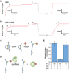
Figure 6. Glutaredoxin reverses the mechanical effects of S-glutathionylation
(A) I9132–75 is S-glutathionylated through cleavage of its disulfide by GSH. Unfolding of I9132–75 increases the protein length by 11 nm, while S-glutathionylation increases it further by 14 nm. Exposure-quench-probe pulse protocol probes the refolding of an I9132–75 polyprotein in the presence of 100 mM GSH. During the exposure pulse, three unfolding events are followed by three S-glutathionylation events (solid arrowheads). The force is then quenched to zero for 5 s. No refolding events are observed during the probe pulse. (B) Recording showing the unfolding and S-glutathionylation of four I9132–75 domains in the presence of 100 mM GSH and 45 µM GRX. After a 5 s quench, refolding is apparent during the probe pulse (25 nm steps, open arrowheads). (C) Diagram of unfolding and S-glutathionylation of I9132–75. Numbers indicate the associated step sizes detected in force-clamp. (D) GRX-mediated deglutathionylation. (E) Refolded fraction after a 5 s quench in 100 mM GSH with or without 45 µM GRX. A baseline for refolding was measured using pre-reduced I9132–75 polyprotein in the presence of 100 mM GSH (N>90). Error bars represent SEM. See also Figure S5.
Figure 7
Figure 7. Modulation of cardiomyocyte elasticity by S-glutathionylation of cryptic cysteines in titin
(A) Single human cardiomyocytes were pulled to different sarcomere lengths and the resulting passive force was measured (Elasticity test). Elasticity was probed initially and after subsequent incubations with 10 mM GSSG, 10 mM GSH, and 1 mM DTT; all incubations were done at long sarcomere lengths (2.6–2.7 µm; Overstretch) to induce unfolding of titin Ig domains. Incubation times are indicated. Top trace shows the stretch protocol; bottom trace show the resulting average passive tension generated by cardiomyocytes. Results are the average of at least 8 cells. Insets show magnification of the relaxation phase after stretching the cardiomyocytes to SL = 2.4 µm. (B) Mean peak force measured at various SLs. GSSG slack is an experiment where incubation with GSSG was done in relaxed cardiomyocytes (SL = 1.8 µm). Forces are normalized to the force measured at 2.4 µm SL in control conditions. Error bars represent SD. Inset: scheme of the experimental setup (C) Model of regulation of muscle elasticity through S-glutathionylation of cryptic cysteines in titin. See also Figure S6.

References

    1. Alegre-Cebollada J, Kosuri P, Rivas-Pardo JA, Fernandez JM. Direct observation of disulfide isomerization in a single protein. Nat Chem. 2011;3:882–887. - PMC - PubMed
    1. Anderson BR, Bogomolovas J, Labeit S, Granzier H. Single molecule force spectroscopy on titin implicates immunoglobulin domain stability as a cardiac disease mechanism. J Biol Chem. 2013;288:5303–5315. - PMC - PubMed
    1. Avner BS, Shioura KM, Scruggs SB, Grachoff M, Geenen DL, Helseth DL, Farjah M, Goldspink PH, Solaro RJ. Myocardial infarction in mice alters sarcomeric function via post-translational protein modification. Mol Cell Biochem. 2012;363:203–215. - PMC - PubMed
    1. Ceyhan-Birsoy O, Agrawal PB, Hidalgo C, Schmitz-Abe K, Dechene ET, Swanson LC, Soemedi R, Vasli N, Iannaccone ST, Shieh PB, et al. Recessive truncating titin gene, TTN, mutations presenting as centronuclear myopathy. Neurology. 2013 - PMC - PubMed
    1. Chen FC, Ogut O. Decline of contractility during ischemia-reperfusion injury: actin glutathionylation and its effect on allosteric interaction with tropomyosin. Am J Physiol Cell Physiol. 2006;290:C719–C727. - PubMed

Publication types

LinkOut - more resources