Review
. 2014 May 6;19(5):741-756.
doi: 10.1016/j.cmet.2014.02.007. Epub 2014 Mar 13.

Central neural regulation of brown adipose tissue thermogenesis and energy expenditure

Affiliations
Review

Central neural regulation of brown adipose tissue thermogenesis and energy expenditure

Shaun F Morrison et al. Cell Metab. .

Abstract

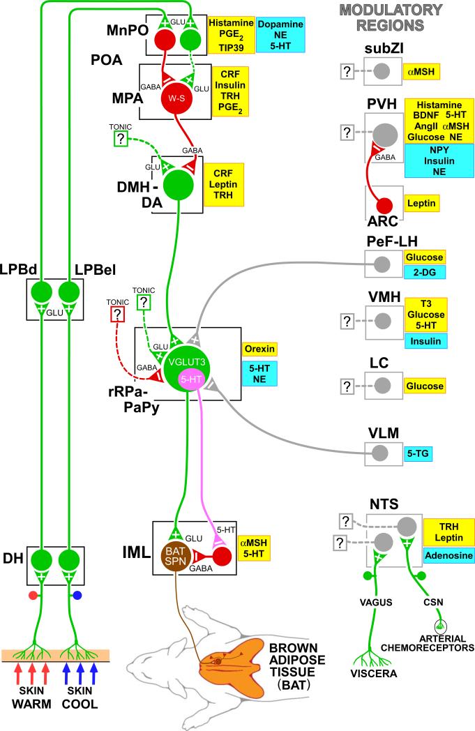
Thermogenesis, the production of heat energy, is the specific, neurally regulated, metabolic function of brown adipose tissue (BAT) and contributes to the maintenance of body temperature during cold exposure and to the elevated core temperature during several behavioral states, including wakefulness, the acute phase response (fever), and stress. BAT energy expenditure requires metabolic fuel availability and contributes to energy balance. This review summarizes the functional organization and neurochemical influences within the CNS networks governing the level of BAT sympathetic nerve activity to produce the thermoregulatory and metabolically driven alterations in BAT thermogenesis and energy expenditure that contribute to overall energy homeostasis.


Figures

Figure 1
Figure 1. Model for the neuroanatomical and neurotransmitter/hormonal organization of the core thermoregulatory network and other CNS sites controlling and modulating brown adipose tissue (BAT) thermogenesis
Cool and warm cutaneous thermal sensory receptors transmit signals to respective primary sensory neurons in the dorsal root ganglia which relay this thermal information to second-order thermal sensory neurons in the dorsal horn (DH). Cool sensory DH neurons glutamatergically activate third-order sensory neurons in the external lateral subnucleus of the lateral parabrachial nucleus (LPBel), while warm sensory DH neurons project to third-order sensory neurons in the dorsal subnucleus of the lateral parabrachial nucleus (LPBd). Thermosensory signals for thermoregulatory responses are transmitted from the LPB to the preoptic area (POA) where GABAergic interneurons in the median preoptic (MnPO) subnucleus are activated by glutamatergic inputs from cool-activated neurons in LPBel and inhibit a BAT-regulating population of warm-sensitive (W-S) neurons in the medial preoptic area (MPA). In contrast, glutamatergic interneurons in the MnPO, postulated to be excited by glutamatergic inputs from warm-activated neurons in LPBd, excite W-S neurons in MPA. Prostaglandin (PG) E2 binds to EP3 receptors to inhibit the activity of W-S neurons in the POA. Preoptic W-S neurons providing thermoregulatory control of BAT thermogenesis inhibit BAT sympathoexcitatory neurons in the dorsomedial hypothalamus and dorsal hypothalamic area (DMH/DA) which, when disinhibited during skin cooling, excite BAT sympathetic premotor neurons in the rostral ventromedial medulla, including the rostral raphe pallidus (rRPa) and parapyramidal area (PaPy), that project to BAT sympathetic preganglionic neurons (SPN) in the spinal intermediolateral nucleus (IML). Some BAT premotor neurons can release glutamate (GLU) to excite BAT sympathetic preganglionic neurons and increase BAT sympathetic nerve activity, while others can release serotonin (5-HT) to interact with 5-HT1A receptors, potentially on inhibitory interneurons in the IML, to increase the BAT sympathetic outflow. Orexinergic neurons in the perifornical lateral hypothalamus (PeF-LH) project to the rRPa to increase the excitability of BAT sympathetic premotor neurons. Activation of neurons in the ventrolateral medulla (VLM) produces an inhibition of BAT thermogenesis, at least in part by noradrenergic activation of α2 receptors on rRPa neurons. Neurochemicals/hormones in yellow boxes activate and those in blue boxes reduce BAT activity. 2-DG, 2-deoxyglucose; 5-HT, 5-hydroxytryptamine; 5-TG, 5-thioglucose; αMSH, alpha melanocyte-stimulating hormone; AngII, angiotensin II; BDNF, brain-derived neurotrophic factor; CRF, corticotrophin releasing factor; CSN, carotid sinus nerve; NE, norepinephrine; NPY, neuropeptide Y; PGE2, prostaglandin E2; T3, triiodothyronine; TIP39, tuberoinfundibular peptide of 39 residues; TRH, thyrotropin-releasing hormone; VGLUT3, vesicular glutamate transporter 3. Copyright 2014 by Oregon Health and Science University.
Figure 2
Figure 2. NTS neurons mediate both inhibition and excitation of BAT thermogenesis
(A) Disinhibitory activation of neurons in intermediate NTS (iNTS) at the level of the area postrema (insert) with injections of bicuculline (BIC) completely reversed the PGE2 in medial preoptic area (MPA)-mediated increases in BAT SNA and BAT temperature (TBAT). Modified from (Cao et al., 2010). (B) Activation of adenosine 1A receptors in iNTS with injections of the agonist, N6-cyclohexyladenosine (CHA), reverses the cooling-evoked increases in BAT SNA and TBAT. Inhibition of iNTS neurons with injections of muscimol (MUSC) reverses the CHA-evoked inhibition of BAT SNA, consistent with adenosine producing an increase in the activity of BAT sympathoinhibitory neurons in iNTS. Modified from (Tupone et al., 2013). (C) Changes in TBAT elicited by agents applied to the 4th ventricle. When preceded by leptin, TRH markedly increases TBAT, indicating BAT thermogenesis. This effect is prevented by prior blockade of signal transduction pathways via 4th ventricle administration of wortmannin to block leptin-evoked PIP3 generation or by the Src-SH2 antagonist, PP2. Modified from (Rogers et al., 2009). (D) Stimulation (400μA, 1 ms pulses, 2 Hz) of afferents in the cervical vagus elicits a potent inhibition of cooling-evoked increases in BAT SNA, TBAT and expired CO2, an indicator of metabolic oxygen consumption. These data reflect a vagal afferent drive to BAT sympathoinhibitory NTS neurons. Author's unpublished observation. (E) Paired shock stimulation (400μA, 1 ms pulses, 6 ms interpulse interval, 0.2 Hz) of afferents in the cervical vagus nerve evokes an excitatory compound action potential in BAT SNA, consistent with a population of vagal afferents activating BAT sympathoexcitatory neurons in NTS. Author's unpublished observation.
Figure 3
Figure 3. Orexinergic and other PeF/LH neurons influence BAT thermogenesis
(A) The activation of BAT SNA and BAT thermogenesis produced by disinhibitory activation of LH neurons with bicuculline (BIC) nanoinjection is dependent on the activation of glutamate receptors on DMH/DA neurons. Modified from (Cerri and Morrison, 2005). (B) Icv PGE2 (filled symbols), but not ACSF (open symbols), elicited a marked increase in BAT temperature in orexin-KO mice (triangles) and in the wild-type littermates for orexin-KO (circles) and for orexin neuron-ablated (squares), but had no effect on TBAT in orexin neuron-ablated mice (diamonds). Modified from (Takahashi et al., 2013). (C) Under cool conditions (TCORE < 37 °C) with a low level of basal BAT SNA, nanoinjection of orexin-A (dashed line) in the rRPa elicited a prolonged increase in BAT SNA and TBAT. Modified from (Tupone et al., 2011). (D) Orexinergic fibers (red) surround putative BAT sympathetic premotor neurons in PaPy (and in rRPa) transynaptically-infected following PRV inoculations of interscapular BAT. Modified from (Tupone et al., 2011).
Figure 4
Figure 4. Paraventricular hypothalamic nucleus (PVH) mechanisms influencing BAT thermogenesis
(A) Histological section through the PVH illustrating the overlap of transynaptically infected, PRV-labeled neurons (brown) following PRV injections into interscapular BAT and in situ hybridization for melanocortin 4-receptor (MC4-R) mRNA expression (black granules). Inset: High magnification of the outlined portion of the PVH. Note the presence of PRV in neurons surrounded by MC4-R (curved black arrows) and PRV in neurons without associated MC4-R (curved open arrows). Bar = 25mm. Modified from (Song et al., 2008). (B) Immunolabeling of PVH neurons for transynaptic infection with PRV (green) after PRV injections into interscapular BAT and for oxytocin (OXY; red). Arrows indicate neurons containing both PRV and OXY (yellow). Modified from (Oldfield et al., 2002). (C) Microinjection of neuropeptide Y (NPY), but not saline vehicle, into the PVH inhibited BAT SNA. Modified from (Egawa et al., 1991). (D) Nanoinjection of bicuculline (BIC) into the PVH completely reversed the cooling-evoked increases in BAT SNA, BAT temperature (TBAT) and expired CO2 (Exp CO2). Modified from (Madden and Morrison, 2009). (E) Schematic of the proposed neurocircuitry mediating influences on BAT thermogenesis mediated by PVH neurons and their GABAergic regulation. Based in part on (Cowley et al., 1999).
Figure 5
Figure 5. Neurons in the ventrolateral medulla (VLM), including catecholaminergic neurons, regulate BAT SNA and BAT thermogenesis
(A) The A1/C1 area of the VLM (bregma −13 mm) contains transynaptically-infected neurons (green) following PRV injections into interscapular BAT, tyrosine hydroxylase (TH)-immunoreactive neurons (red), and double-labeled (yellow, arrowhead) neurons. (B) Following PRSx8-channel rhodopsin 2-mCherry (ChR2) lentivirus nanoinjections into VLM, many dopamine beta hydroxylase (DβH)-expressing neurons (green) are double-labeled (yellow, arrowheads) due to transfection with ChR2 (red). (C) Following PRSx8-channel rhodopsin 2-mCherry (ChR2) lentivirus nanoinjections into VLM, the rRPa (white dotted outline) contains highly varicose fibers (red) expressing ChR2. (D) Laser photostimulation of VLM neurons containing the ChR2 (largely catecholaminergic neurons) inhibited BAT SNA and reduced TBAT, an effect that was attenuated by blockade of α2-adrenergic receptors in the rRPa. Panels A – D, suggesting that activation of catecholaminergic neurons in the VLM inhibits BAT SNA via direct catecholaminergic inputs to the rostral raphe pallidus (rRPa), are modified from (Madden et al., 2013). (E) Glucoprivation (2-DG, iv) activates (increases c-fos (black nuclei)) many catecholaminergic neurons (gray) in the A1/C1 area of the VLM. Modified from (Ritter et al., 1998). (F) Local glucoprivation in the VLM by nanoinjection of 5-thioglucose (5-TG, dashed line) completely inhibits BAT SNA and reduces BAT thermogenesis (TBAT) and metabolic oxygen consumption (EXP CO2). Modified from (Madden, 2012).

References

    1. Acheson K, Jequier E, Wahren J. Influence of beta-adrenergic blockade on glucose-induced thermogenesis in man. J. Clin. Invest. 1983;72:981–986. - PMC - PubMed
    1. Almeida MC, Steiner AA, Coimbra NC, Branco LG. Thermoeffector neuronal pathways in fever: a study in rats showing a new role of the locus coeruleus. J. Physiol. 2004;558:283–294. - PMC - PubMed
    1. Amini-Sereshki L, Zarrindast MR. Brain stem tonic inhibition of thermoregulation in the rat. Am. J. Physiol. Regul. Integr. Comp. Physiol. 1984;247:R154–159. - PubMed
    1. Arruda AP, Milanski M, Romanatto T, Solon C, Coope A, Alberici LC, Festuccia WT, Hirabara SM, Ropelle E, Curi R, Carvalheira JB, Vercesi AE, Velloso LA. Hypothalamic actions of tumor necrosis factor alpha provide the thermogenic core for the wastage syndrome in cachexia. Endocrinology. 2010;151:683–694. - PubMed
    1. Bachman ES, Dhillon H, Zhang CY, Cinti S, Bianco AC, Kobilka BK, Lowell BB. betaAR signaling required for diet-induced thermogenesis and obesity resistance. Science. 2002;297:843–845. - PubMed

Publication types

MeSH terms

LinkOut - more resources