. 2014 Mar 27;6(6):1017-1025.
doi: 10.1016/j.celrep.2014.01.043. Epub 2014 Mar 13.

The Marburg virus VP24 protein interacts with Keap1 to activate the cytoprotective antioxidant response pathway

Affiliations

The Marburg virus VP24 protein interacts with Keap1 to activate the cytoprotective antioxidant response pathway

Megan R Edwards et al. Cell Rep. .

Abstract

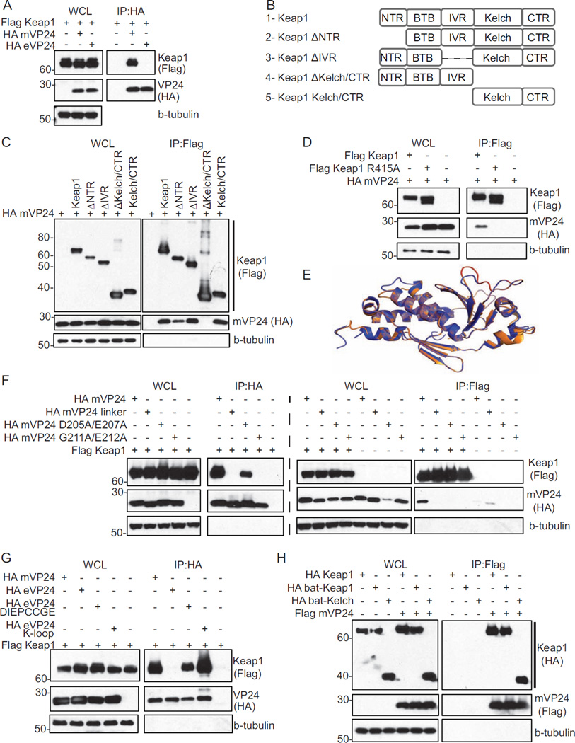
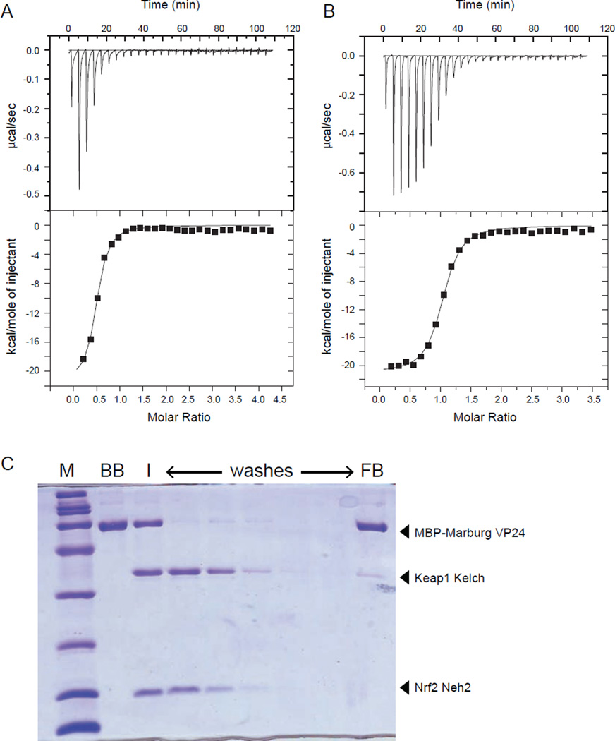
Kelch-like ECH-associated protein 1 (Keap1) is a ubiquitin E3 ligase specificity factor that targets transcription factor nuclear factor (erythroid-derived 2)-like 2 (Nrf2) for ubiquitination and degradation. Disrupting Keap1-Nrf2 interaction stabilizes Nrf2, resulting in Nrf2 nuclear accumulation, binding to antioxidant response elements (AREs), and transcription of cytoprotective genes. Marburg virus (MARV) is a zoonotic pathogen that likely uses bats as reservoir hosts. We demonstrate that MARV protein VP24 (mVP24) binds the Kelch domain of either human or bat Keap1. This binding is of high affinity and 1:1 stoichiometry and activates Nrf2. Modeling based on the Zaire ebolavirus (EBOV) VP24 (eVP24) structure identified in mVP24 an acidic loop (K-loop) critical for Keap1 interaction. Transfer of the K-loop to eVP24, which otherwise does not bind Keap1, confers Keap1 binding and Nrf2 activation, and infection by MARV, but not EBOV, activates ARE gene expression. Therefore, MARV targets Keap1 to activate Nrf2-induced cytoprotective responses during infection.


Figures

Fig. 1
Fig. 1. mVP24 interacts with Keap1 in co-immunoprecipitation assays
(A) Co-immunoprecipitations (co-IP) with anti-HA antibody were performed on lysates of HEK293T cells co-transfected with plasmids for Flag-Keap1 and HA-mVP24 or HA-eVP24. Western blots were performed for Flag and HA. (B) Schematic diagram of Flag tagged Keap1 domain deletion mutants used in (C). (C) Flag-Keap1 domain deletion mutant constructs were co-expressed in HEK293T cells with HA-mVP24 and analyzed by co-IP with anti-Flag antibody. (D) HA-mVP24 and either Flag-Keap1 or Flag-Keap1 R415A were analyzed by co-IP as in (C). (E) Overlay of the mVP24 structural model (orange) on the determined eVP24 structure (purple). The mVP24 K-loop (amino acids 205–212) is indicated in red. (F) Flag-Keap1 and HA-mVP24 wild-type or mutants were analyzed by co-IP as in (A) and (C). (G) Flag-Keap1 and HA-mVP24, eVP24, eVP24 DIEPCCGE or eVP24 K-loop were co-expressed in HEK293T cells and analyzed by co-IP as in (A). (H) Flag-mVP24 and HA-Keap1, bat-Keap1 and bat-Kelch were co-expressed in HEK293T cells and analyzed by co-IP as in (C). See also Figure S1.
Fig. 2
Fig. 2. Marburg VP24 binds to Keap1 Kelch domain with high affinity and specificity
mVP24 binds to Keap1 Kelch domain with high affinity and specificity. (A and B) Representative ITC data for KELCH domain of Keap1 binding to (A) Nrf2 Neh2 domain and (B) mVP24. Raw heats of reaction vs. time (top panels) and the integrated heats of reaction vs. molar ratio of ligand to receptor (bottom panels) are shown. Thermodynamic binding parameters of KD = 170 ± 60 nM, ΔH = −1.96 ± 0.1×10⁴ kcal/mol, TΔS = −10.4 kcal/mol, and n (no. of sites) = 0.49 ± 0.02 for (A) and KD = 158 ± 20 nM, ΔH = −2.10 ± 0.03× 10⁴ kcal/mol, TΔS = −11.7 kcal/mol, and n (no. of sites) = 1.00 ± 0.01 for (B) were obtained. (C) mVP24 binding to Kelch prevents Nrf2 Neh2 interaction. Coomassie-blue stained SDS-PAGE of a pulldown assay where MBP-mVP24 was immobilized on amylose resin (BB, bound beads). Keap1 Kelch and Nrf2 Neh2 domain was subsequently added to the resin (I, input), and the resin was washed with buffer (washes). The final bound bead sample (FB, final beads) is indicated. See also Figure S2.
Fig. 3
Fig. 3. mVP24 activates expression of ARE genes
(A and B) HEK293T cells were transfected with the ARE luciferase reporter plasmid, a constitutively expressed Renilla luciferase plasmid, and pCAGGS (empty vector) or increasing concentrations of HA-p62, Flag-wild-type mVP24 or mVP24 mutants. (B) Same as (A), with the additional over-expression of Flag-Nrf2 and Flag-Keap1. At 18 hours post transfection (hpt) luciferase activity was assayed for (A) and (B). Western blots performed for HA and Flag are indicated. (C) Same assay protocol as (A) but transfected with HA-mVP24, eVP24 or eVP24 mutants. (D) pCAGGS, Flag-Nrf2, mVP24 or mVP24 G211A/E212A were transfected in triplicate in HEK293T cells. At 24 hpt, qRT-PCR was performed to quantify mRNAs for the indicated genes, normalized to the RPS11 mRNA. (E) HEK293T cells were transfected with the indicated plasmids and 18hpt endogenous NQO1 was measured by western blot. (F) Cell viability assay. HEK293T cells were transfected with pCAGGS, Flag-Nrf2, mVP24 or mVP24 G211A/E212A and 24 hpt were treated with vehicle control (ethanol) or 5uM menadione (M) for three hours. (A-D) Represent the mean and SEM of triplicate samples and statistical significance was assessed by a one-way ANOVA comparing columns to the control (white bar), ***p<0.001, **p<0.01 and *p<0.05. Samples in (F) represent the mean and SEM of six samples and significance was assessed by a one-way ANOVA, *p<0.05. See also Figure S3.
Fig. 4
Fig. 4. MARV infection upregulates the Nrf2 antioxidant pathway
(A and B) THP-1 cells were infected with MARV-Ang or Zaire EBOV (MOI=3) and subjected to expression analysis by mRNA-seq. (A) Heat map displaying the expression profile of 30 Nrf2-activated genes (Chorley et al., 2012)). Red indicates up-regulated genes (max induction = 8.55 fold relative to mock-infected cells). Green indicates down-regulated genes (lowest value=0.2 fold relative to mock-infected cells). Gray indicates genes undetected in the mRNA-seq. (B) mVP24 and eVP24 mRNA expression levels represented as median nucleotide coverage. (C) THP-1 cells were infected with MARV-Ang or MARV-Mus (MOI=1) and subjected to qRT-PCR. Values were normalized to RPS11. Mock sample contains a single replicate; MARV-Ang and MARV-Mus represent the mean and SEM of triplicate samples. See also Figure S4.

Comment in

References

    1. Amman BR, Carroll SA, Reed ZD, Sealy TK, Balinandi S, Swanepoel R, Kemp A, Erickson BR, Comer JA, Campbell S, et al. Seasonal pulses of Marburg virus circulation in juvenile Rousettus aegyptiacus bats coincide with periods of increased risk of human infection. PLoS Pathog. 2012;8:e1002877. - PMC - PubMed
    1. Baird L, Dinkova-Kostova AT. The cytoprotective role of the Keap1-Nrf2 pathway. Arch Toxicol. 2011;85:241–272. - PubMed
    1. Bamberg S, Kolesnikova L, Moller P, Klenk HD, Becker S. VP24 of Marburg virus influences formation of infectious particles. J Virol. 2005;79:13421–13433. - PMC - PubMed
    1. Beniac DR, Melito PL, Devarennes SL, Hiebert SL, Rabb MJ, Lamboo LL, Jones SM, Booth TF. The organisation of Ebola virus reveals a capacity for extensive, modular polyploidy. PLoS One. 2012;7:e29608. - PMC - PubMed
    1. Bharat TA, Noda T, Riches JD, Kraehling V, Kolesnikova L, Becker S, Kawaoka Y, Briggs JA. Structural dissection of Ebola virus and its assembly determinants using cryo-electron tomography. Proc Natl Acad Sci U S A. 2012;109:4275–4280. - PMC - PubMed

Publication types

MeSH terms