. 2014 Mar 31;24(7):725-30.
doi: 10.1016/j.cub.2013.12.051. Epub 2014 Mar 13.

Sexually dimorphic octopaminergic neurons modulate female postmating behaviors in Drosophila

Affiliations

Sexually dimorphic octopaminergic neurons modulate female postmating behaviors in Drosophila

Carolina Rezával et al. Curr Biol. .

Abstract

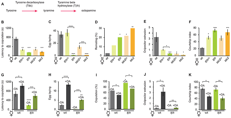
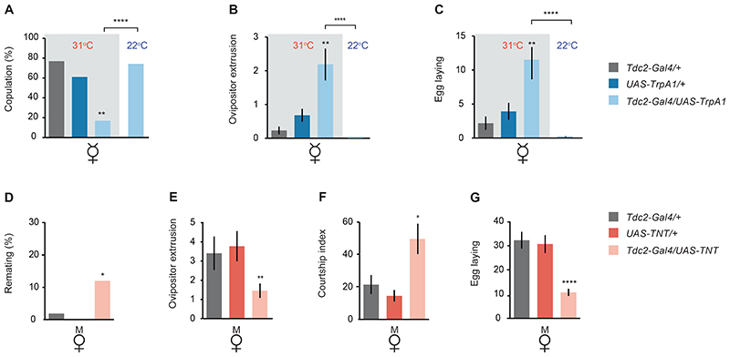
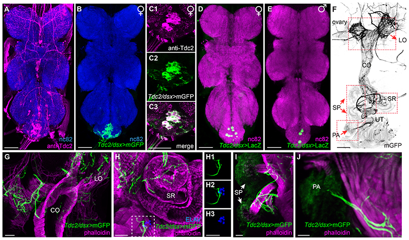
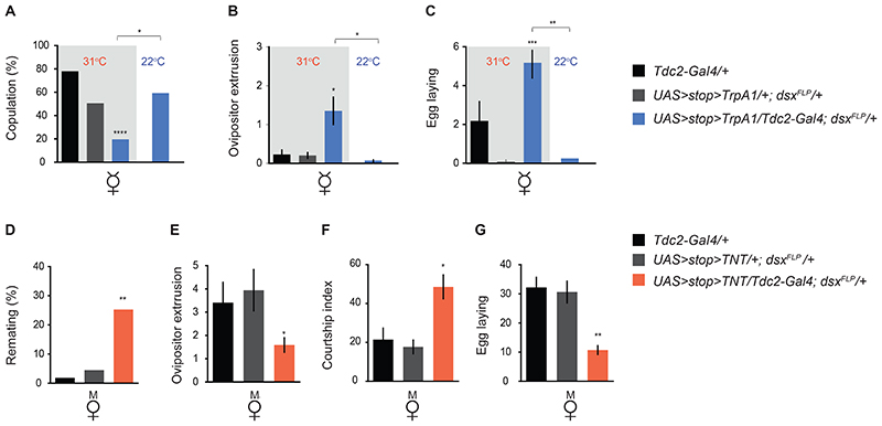
Mating elicits profound behavioral and physiological changes in many species that are crucial for reproductive success. After copulation, Drosophila melanogaster females reduce their sexual receptivity and increase egg laying [1, 2]. Transfer of male sex peptide (SP) during copulation mediates these postmating responses [1, 3-6] via SP sensory neurons in the uterus defined by coexpression of the proprioceptive neuronal marker pickpocket (ppk) and the sex-determination genes doublesex (dsx) and fruitless (fru) [7-9]. Although neurons expressing dsx downstream of SP signaling have been shown to regulate postmating behaviors [9], how the female nervous system coordinates the change from pre- to postcopulatory states is unknown. Here, we show a role of the neuromodulator octopamine (OA) in the female postmating response. Lack of OA disrupts postmating responses in mated females, while increase of OA induces postmating responses in virgin females. Using a novel dsx(FLP) allele, we uncovered dsx neuronal elements associated with OA signaling involved in modulation of postmating responses. We identified a small subset of sexually dimorphic OA/dsx(+) neurons (approximately nine cells in females) in the abdominal ganglion. Our results are consistent with a model whereby OA neuronal signaling increases after copulation, which in turn modulates changes in female behavior and physiology in response to reproductive state.


Figures

Figure 1
Figure 1. Octopamine regulates female receptivity and postmating responses.
(A) The OA biosynthesis cascade. (B) Lack of OA increases receptivity in virgin females. Tβh (TβhnM18) and Tdc2 (Tdc2RO54) mutant virgin females show increased receptivity, measured as latency to copulation (in seconds); n = 25-35. (C-F) Lack of OA disrupts postmating responses in mated females. Tβh (TβhnM18) and Tdc2 (Tdc2RO54) mutant mated females show disrupted postmating responses. (C) Number of eggs laid per female 48 hr after copulation; n = 20-35. (D) Remating frequency for females tested 48 hr after the initial mating; n = 40-60. (E) Female ovipositor extrusion per minute during courtship; n =18-35. (F) Male courtship index of wild-type males paired with females of the indicated genotypes; n = 15-25. (G-K) Increasing OA levels reduces receptivity and triggers postmating responses in wild-type and Tβh (TβhnM18) mutant virgin females. (G) Latency to copulation (in seconds); n = 30-40. (H) Number of eggs laid per female after 6 days; n = 45-55. (I) Percentage of females that copulated within 1 hr; n = 35-45. (J) Female ovipositor extrusion per minute during courtship; n= 16-20. (K) Male courtship index of wild-type males paired with females of the indicated genotypes; n = 18-22. CS flies were used as wild-type in all behavioral tests. Females labeled +OA were fed 7.5 mg/mL of OA. Error bars indicate SEM. Statistical comparisons of the indicated genotypes were made against CS (B-F) or OA- wild-type controls (G-K), unless otherwise indicated. A Kruskal-Wallis ANOVA test was performed in B,C,E-H and J,K, and Fisher exact test in D, I. *p<0.05, **p<0.01, ***p<0.001, ****p<0.0001. Virgin and mated females are indicated as V and M, respectively.
Figure 2
Figure 2. Tdc2+ neurons are involved in postmating behaviors.
(A-C) Artificially activating Tdc2+ neurons decreases receptivity in virgin females. (A) Percentage of females that copulated within 1 hr; n = 35-45. (B) Female ovipositor extrusion per minute during courtship; n = 25-30. (C) Number of eggs laid per female during 48 hr; n = 35-45. (D-G) Silencing Tdc2+ neurons disrupts postmating responses in mated females. (D) Remating frequency for females tested 48 hr after the initial mating; n = 30-40. (E) Female ovipositor extrusion per minute during courtship; n = 12-16. (F) Male courtship index of wild-type males paired with females of the indicated genotypes; n = 15-18. (G) Number of eggs laid per female 48 hr after copulation; n = 25-35. Error bars indicate ±SEM. Statistical comparisons were made against Tdc2-Gal4/+ (A-G) and UAS-TrpA1/+ in (A-C) or UAS-TNT/+ (in D-G), unless otherwise indicated. Kruskal-Wallis ANOVA test was performed in B,C,E-G and Fisher exact test in A,D. *p<0.05, **p<0.01, ***p<0.001, ****p<0.0001. Virgin and mated females are indicated as V and M, respectively. Note that for Tdc2-Gal4/UAS-Trpa1 at 22°C in (B-C) and for UAS-TNT/+ in (D), error bars are not visible as they are close to 0.
Figure 3
Figure 3. Identification of Tdc2+/dsx+ co-expressing neurons in the nervous system.
Combination of the dsxFLP line with Tdc2-Gal4 allows expression of Gal4/FLP-responsive UAS>stop>reporters in Tdc2/dsx+-coexpressing neurons. (A-C) Visualization of Tdc2+ and Tdc2/dsx+ cell bodies and projections in the female VNC. Tdc2+ neurons are visualized with anti-Tdc2 antibody (magenta) in the female VNC (A). Approximately nine Tdc2/dsx+ cell bodies are labeled with UAS>stop>mCD8::GFP reporter (Tdc2/dsx>mGFP; green) in the female VNC (B and C). Higher magnification of the female Abg in (B) depicting Tdc2/dsx+ neurons (green) colabeled with anti-Tdc2 antibody (magenta) is shown in (C1)-(C3). In (A) and (B), neuropil is counterstained with anti-nC82 (blue). (D and E) Visualization of Tdc2/dsx+ neuronal nuclei expressing the UAS>stop>nLacZ reporter (Tdc2/dsx>LacZ; green). Approximately nine Tdc2/dsx+ nuclei are detected in the female VNC (D) and approximately three in the male VNC (E). Anti β-Gal is shown in green. Neuropil is counterstained with anti-nC82 (magenta). (F-J) Visualization of Tdc2/dsx+ cell bodies and projections in the female reproductive system. (F) Female reproductive system showing Tdc2/dsx+ innervations (Tdc2/dsx > mGFP; black) in the lateral oviducts (LO), common oviduct (CO), uterus (UT), spermathecae (SP), seminal receptacle (SR), and parovaria (PA) (indicated by red arrows). (G-J) Higher magnification of lateral and common oviducts (G), seminal receptacle (H), spermathecae (I), and parovaria (J). Tdc2/dsx+ neuronal projections are shown in green (Tdc2/dsx>mGFP) and phalloidin (a marker for F-actin) in magenta. Lack of colocalization between Tdc2/dsx+ neuronal cell bodies and SP sensory nuclei (white box in H) is shown at higher magnification in (H1)-(H3). Tdc2/dsx+ neurons are shown in green, and SP sensory neurons are stained with the neuronal nuclear marker anti-ELAV (blue). Scale bars represent 50 μm (A, B, D, and E), 100 μm (F), and 25 μm (C and G−J). See also Figures S2 and S3.
Figure 4
Figure 4. A subset of sexually dimorphic Tdc2/dsx+ neurons is required for female postmating behavioral responses.
Combination of the dsxFLP line with Tdc2-Gal4 allows expression of the selected effectors, UAS>stop>TrpA1 (A-C) or UAS>stop>TNT (D-G) in all intersecting neurons (dsx⋂Tdc2). (A-C) Artificial activation of Tdc2/dsx+ neurons reduces receptivity and increases postmating responses in virgin females. (A) Receptivity was scored as percentage of females that copulated within 1 hr. n = 45-55. (B) Female ovipositor extrusion per minute during courtship. n = 23-33. (C) Number of eggs laid per female in 48 hr after copulation. n = 25-45. (D-G) Silencing of Tdc2/dsx+ neurons reduces postmating responses in mated females. (D) Remating frequency for females tested 48 hr after the initial mating. n = 30-40. (E) Female ovipositor extrusion per minute during courtship. n = 14-24. (F) Male courtship index of wild-type males paired with females of the indicated genotypes. n = 35-45. (G) Number of eggs laid per female 48 hr after copulation. n = 25-35. Error bars indicate ± SEM. A Kruskal-Wallis ANOVA test was performed in B,C and E-G and Fisher’s exact test in A,D. Statistical comparisons were made against Tdc2-Gal4/+ (A-G) and UAS>stop>TrpA1/+;dsxFLP/+ (A−C) or UAS>stop>TNT/+;dsxFLP/+ (D-G), unless otherwise indicated. *p < 0.05, **p < 0.01, and ***p < 0.001. Virgin and mated females are indicated as V and M, respectively.

Comment in

References

    1. Kubli E. Sex-peptides: seminal peptides of the Drosophila male. Cell Mol Life Sci. 2003;60:1689–1704. - PMC - PubMed
    1. Avila FW, Sirot LK, LaFlamme BA, Rubinstein CD, Wolfner MF. Insect seminal fluid proteins: identification and function. Annu Rev Entomol. 2011;56:21–40. - PMC - PubMed
    1. Chen PS, Stumm-Zollinger E, Aigaki T, Balmer J, Bienz M, Bohlen P. A male accessory gland peptide that regulates reproductive behavior of female D. melanogaster. Cell. 1988;54:291–298. - PubMed
    1. Aigaki T, Fleischmann I, Chen PS, Kubli E. Ectopic expression of sex peptide alters reproductive behavior of female D. melanogaster. Neuron. 1991;7:557–563. - PubMed
    1. Liu H, Kubli E. Sex-peptide is the molecular basis of the sperm effect in Drosophila melanogaster. Proc Natl Acad Sci U S A. 2003;100:9929–9933. - PMC - PubMed

Publication types

Associated data

LinkOut - more resources