. 2014 May:42:12-23.
doi: 10.1016/j.neuro.2014.02.009. Epub 2014 Mar 11.

Quantitative mapping of trimethyltin injury in the rat brain using magnetic resonance histology

Affiliations

Quantitative mapping of trimethyltin injury in the rat brain using magnetic resonance histology

G Allan Johnson et al. Neurotoxicology. 2014 May.

Abstract

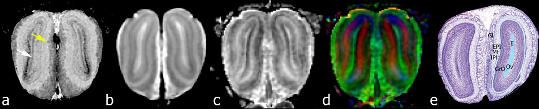
The growing exposure to chemicals in our environment and the increasing concern over their impact on health have elevated the need for new methods for surveying the detrimental effects of these compounds. Today's gold standard for assessing the effects of toxicants on the brain is based on hematoxylin and eosin (H&E)-stained histology, sometimes accompanied by special stains or immunohistochemistry for neural processes and myelin. This approach is time-consuming and is usually limited to a fraction of the total brain volume. We demonstrate that magnetic resonance histology (MRH) can be used for quantitatively assessing the effects of central nervous system toxicants in rat models. We show that subtle and sparse changes to brain structure can be detected using magnetic resonance histology, and correspond to some of the locations in which lesions are found by traditional pathological examination. We report for the first time diffusion tensor image-based detection of changes in white matter regions, including fimbria and corpus callosum, in the brains of rats exposed to 8 mg/kg and 12 mg/kg trimethyltin. Besides detecting brain-wide changes, magnetic resonance histology provides a quantitative assessment of dose-dependent effects. These effects can be found in different magnetic resonance contrast mechanisms, providing multivariate biomarkers for the same spatial location. In this study, deformation-based morphometry detected areas where previous studies have detected cell loss, while voxel-wise analyses of diffusion tensor parameters revealed microstructural changes due to such things as cellular swelling, apoptosis, and inflammation. Magnetic resonance histology brings a valuable addition to pathology with the ability to generate brain-wide quantitative parametric maps for markers of toxic insults in the rodent brain.

Keywords: Animal models; Environmental toxins; MRI; Rat; Trimethyltin.


Figures

Fig. 1
Fig. 1
Three image-processing pipelines were constructed to facilitate processing of the multiple three-dimensional arrays and to ensure that the data were all processed in identical fashion. Software modules on multiple computers are connected via Perl scripts to automate the steps within and between the pipelines, including: image reconstruction, diffusion tensor estimation, and image registration. Image registration consists of both affine and diffeomorphic mapping (diff morph).
Fig. 2
Fig. 2
Representative images are shown from a control animal in a dorsal plane: (a) GRE image; (b) diffusion weighted image (DWI); (c) fractional anisotropy (FA) image; (d) color fractional anisotropy (clrFA). Py (pyramidal cell layer); CA1, CA2, CA3 (cornu ammonis fields of the hippocampus); GrDG (granular layers of the dentate gyrus); CPu (caudate putamen [fibers] – see arrow).
Fig. 3
Fig. 3
Coronal slices from the same data as in Fig. 2 demonstrate the neuroanatomy in the olfactory bulb in (a) GRE image, where cell layers can be seen, e.g. the ependymal layer (white arrow), as well as glomeruli (yellow arrow) in the outer layer; (b) diffusion weighted image (DWI); (c) fractional anisotropy (FA) image; (d) color fractional anisotropy (clrFA); (e) labeled cell layers in top of H&E stain from similar slice through the rat brain atlas (adapted from and reprinted with permission from Elsevier Academic Press, San Diego, Plate 1 in: George Paxinos and Charles Watson, The Rat Brain in Stereotaxic Coordinates 6th ed., ISBN-13 978-0125476126, 2007). E (ependymal layer); Ov (olfactory ventricle); Gl (glomerular layer); EPl (external plexiform layer); IPl (internal plexiform layer); MI (mitral cells layer); GrO (granule cells layer of olfactory).
Fig. 4
Fig. 4
Sagittal slice through a control rat brain showing (a) diffusion weighted image (DWI), and (b) the minimum deformation template obtained from the 8 diffusion weighted images from the control specimens (inset shows DWI through the hippocampus, magnified in Fig. 5b). Notice white matter striations (arrows) within the caudate putamen (CPu) in the single specimen (a), relative to the minimum deformation template (b).
Fig. 5
Fig. 5
Comparison of (a) conventional AChE histology from Paxinos and Watson atlas (2007) and (b) diffusion weighted image (DWI) through the hippocampus (see inset in Fig. 4b), with (c) labels of cellular layers in the Paxinos and Watson atlas demonstrate how well MR delineates the boundaries in many of these subtle neuroanatomical structures (arrow in Fig. 5b) (adapted from and reprinted with permission from Elsevier Academic Press, San Diego, Plate 169 in: George Paxinos and Charles Watson, The Rat Brain in Stereotaxic Coordinates 6th ed., ISBN-13 978–0125476126, 2007). The superior colliculus layers depicted include: (1) SuG (superficial gray); (2) Op (optic nerve layer); (3) InG (internal gray layer); (4) InWh (internal white layer); as well as immediately ventral the DpG (deep gray layer). The hippocampal layers are also clearly visible: Or (oriens); Rad (stratum radiatum); LMol (lacunosum moleculare); MoDG (molecular layer of dentate gyrus); DG (dentate gyrus); Hil (hilus).
Fig. 6
Fig. 6
The corrected p values (Q) are superimposed on the average diffusion weighted image from the control group demonstrating areas where there is morphologic difference between the control and 8mg/Kg-dose groups (column 1) and control and 12 mg/Kg-dose group (column 2). A potential confound we need to acknowledge is due to the 3 days difference in the time point evaluation, where for humane reasons, rats on 12 mg/Kg were sacrificed earlier than rats in the 8 mg/Kg-dose cohort. Regions in which there are differences in the FA are shown for comparison between the control and low-dose (column 3) and control and high-dose group (column 4). White arrows indicate anatomical regions identified by H&E as different in TMT-treated rats.
Fig. 7
Fig. 7
H&E sections through: a) the olfactory bulb; b) piriform cortex; c) hippocampal dentate gyrus; d) substantia nigra; e) red nucleus, and f) cerebellum show widely dispersed evidence of necrosis (yellow arrows) and neuronal central chromatolysis (black arrows) in 12 mg/Kg TMT-treated rats. White arrows on MRH images indicate the approximate location for the H&E section details on the magnetic resonance histology reference atlas (Johnson et al. 2012).
Fig. 8
Fig. 8
Statistical maps showing the effect size for local volume changes and FA parameters (FDR 0.15) between the two TMT-treated animal cohorts, relative to controls. Effect sizes are attributed to TMT dose, but we should acknowledge a difference of 3 days in time points for the 12 mg/Kg-group. Effect sizes are assigned into one of four categories, from low (<10%), to mild (10–30%), medium (30–50%), and large (>50%) effects. The same coronal sections as in Fig. 6 examined for local changes in DTI parametric maps after correcting for morphometric changes. Regions with significant differences in fractional anisotropy (FA) between TMT-treated rats and saline injected controls include: (1) granule cells layer in the olfactory bulbs; (2) the piriform cortex and striatum, as well as external capsule; (3) dentate gyrus of hippocampus (well defined in the 12mg/Kg-dose treated group), cerebellar nuclei, and white matter.

References

    1. Avants BB, Epstein CL, Grossman M, Gee JC. Symmetric diffeomorphic image registration with cross-correlation: evaluating automated labeling of elderly and neurodegenerative brain. Med Image Anal. 2008;12(1):26–41. - PMC - PubMed
    1. Badea A, Gewalt S, Avants BB, Cook JJ, Johnson GA. Quantitative mouse brain phenotyping based on single and multispectral MR protocols. Neuroimage. 2012;63(3):1633–1645. http://dx.doi.org/10.1016/j.neuroimage.2012.07.021. - DOI - PMC - PubMed
    1. Badea A, Johnson GA, Williams RW. Genetic dissection of the mouse brain using high-field magnetic resonance microscopy. Neuroimage. 2009;45(4):1067–1079. http://dx.doi.org/10.1016/j.neuroimage.2009.01.021. - DOI - PMC - PubMed
    1. Badea A, Kostopoulos GK, Ioannides AA. Surface visualization of electromagnetic brain activity. J Neurosci Methods. 2003;127(2):137–147. http://dx.doi.org/10.1016/S0165-0270(03)00100-6. - DOI - PubMed
    1. Badea A, Williams RW, Johnson GA. Proceedings of Society for Neuroscience. San Diego, CA: 2007. Nov, Magnetic resonance microscopy-based brain morphometry in BXD recombinant inbred mice; pp. 3–7.

Publication types

LinkOut - more resources