. 2014 Jun;32(5):541-50.
doi: 10.1016/j.mri.2014.02.002. Epub 2014 Feb 10.

XeNA: an automated 'open-source' (129)Xe hyperpolarizer for clinical use

Affiliations

XeNA: an automated 'open-source' (129)Xe hyperpolarizer for clinical use

Panayiotis Nikolaou et al. Magn Reson Imaging. 2014 Jun.

Abstract

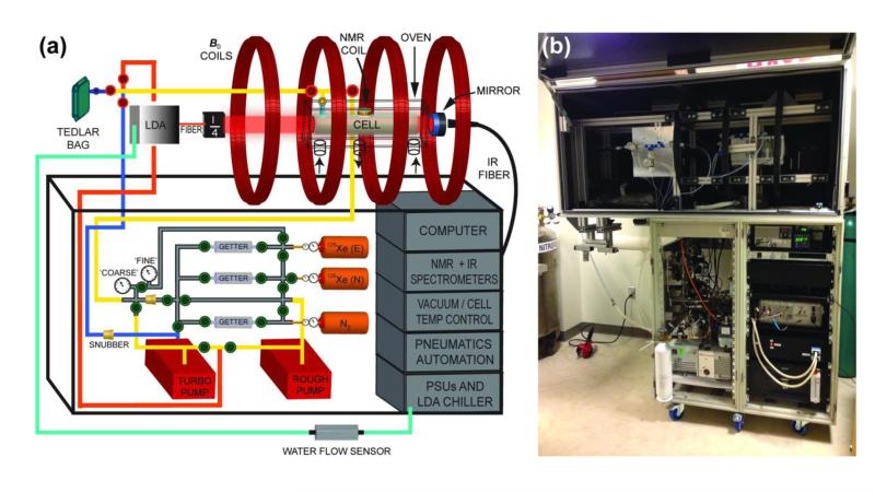
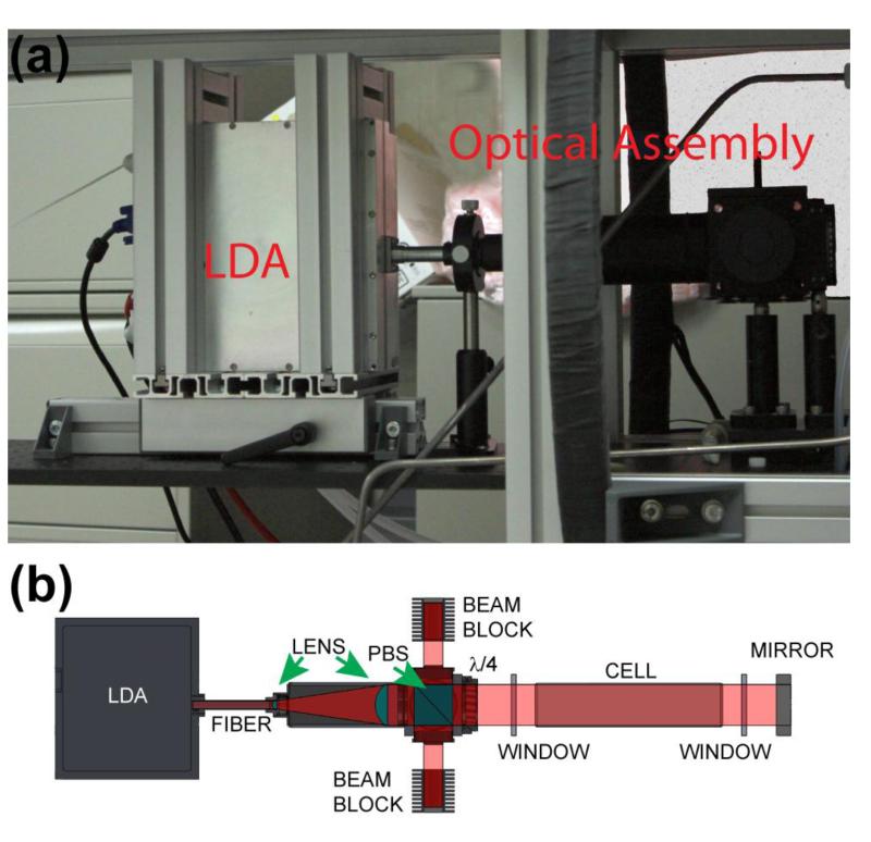
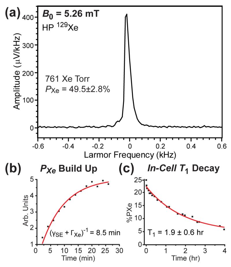
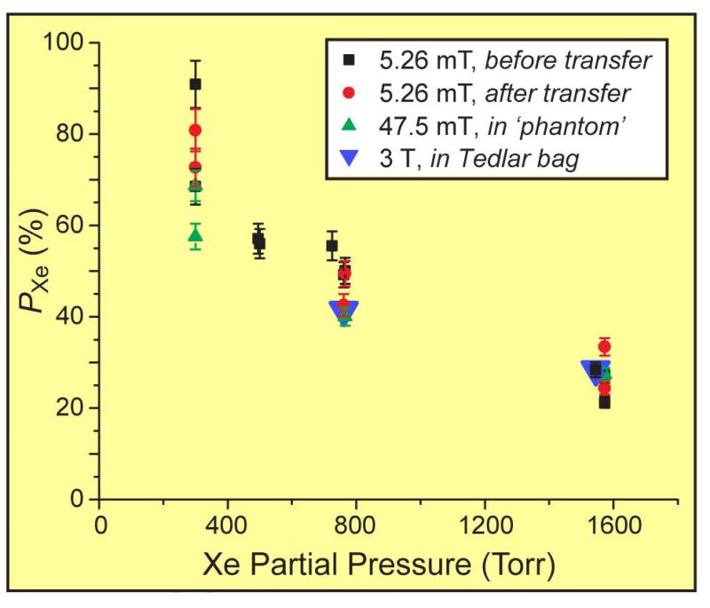
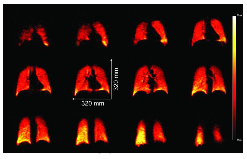
Here we provide a full report on the construction, components, and capabilities of our consortium's "open-source" large-scale (~1L/h) (129)Xe hyperpolarizer for clinical, pre-clinical, and materials NMR/MRI (Nikolaou et al., Proc. Natl. Acad. Sci. USA, 110, 14150 (2013)). The 'hyperpolarizer' is automated and built mostly of off-the-shelf components; moreover, it is designed to be cost-effective and installed in both research laboratories and clinical settings with materials costing less than $125,000. The device runs in the xenon-rich regime (up to 1800Torr Xe in 0.5L) in either stopped-flow or single-batch mode-making cryo-collection of the hyperpolarized gas unnecessary for many applications. In-cell (129)Xe nuclear spin polarization values of ~30%-90% have been measured for Xe loadings of ~300-1600Torr. Typical (129)Xe polarization build-up and T1 relaxation time constants were ~8.5min and ~1.9h respectively under our spin-exchange optical pumping conditions; such ratios, combined with near-unity Rb electron spin polarizations enabled by the high resonant laser power (up to ~200W), permit such high PXe values to be achieved despite the high in-cell Xe densities. Importantly, most of the polarization is maintained during efficient HP gas transfer to other containers, and ultra-long (129)Xe relaxation times (up to nearly 6h) were observed in Tedlar bags following transport to a clinical 3T scanner for MR spectroscopy and imaging as a prelude to in vivo experiments. The device has received FDA IND approval for a clinical study of chronic obstructive pulmonary disease subjects. The primary focus of this paper is on the technical/engineering development of the polarizer, with the explicit goals of facilitating the adaptation of design features and operative modes into other laboratories, and of spurring the further advancement of HP-gas MR applications in biomedicine.

Keywords: Hyperpolarization; Laser-polarized xenon; Lung imaging; MRI; Optical pumping.


Figures

Figure 1
Figure 1
The XeNA polarizer. (a) of the polarizer’s key components (the self-pressurized liquid N2 dewar that provides gas for heating and cooling of the oven and N2 gas cylinder used to operate pneumatic valves are not shown) (40). The optical path is represented by (“λ/4”) and is comprised of beam expanding optics, polarizing beam-splitter cube, quarter-wave plate, and heat sinks (see Fig. 2). For the gas cylinders, “N” and “E” designate xenon with naturally-abundant 129Xe and isotopically-enriched 129Xe, respectively (40). (b) Photograph of XeNA with open laser enclosure in its current location in a clinical MRI suite at Brigham and Womens’ Hospital, Boston, MA, USA.
Figure 2
Figure 2
(a) Laser diode array with translational mounting frame, and optical path assembly. (b) Schematic showing the principal elements of the optical path (40). The beam blocks are drawn separated from the polarizing beam splitter (PBS) housing for clarity.
Figure 3
Figure 3
(a) OP-Oven with OP-Cell (Mid-Rivers Glassblowing, Inc., St. Charles, MO, P/N MRG934-01A) mounted inside, with gas manifold components shown connected to the OP-Cell. (b) Glass (Pyrex) spiral storage condenser (Mid-Rivers Glassblowing, Inc., St. Charles, MO, P/N MRG927-01C) controlled via helical-rotary actuator assembly, residing in a strong (>500 Gauss) magnetic field produced by a pair of large 4 in. × 4 in. × 1 in. neodymium-iron-boride permanent magnets (Indigo Instruments). The magnet yoke was custom-machined from aluminum. (c) Two 400 W T-Type heaters (Omega Engineering, Stamford, CT) mounted inside aluminum enclosure. (d) Custom-made Helical connected and Humphrey Rotary Actuator assembly allowing automation of the Chem-Glass Teflon stopcocks. A video of the glass-valve actuation procedure can be viewed at:http://www.youtube.com/watch?v=w33xs9KHuB0.
Figure 4
Figure 4
(a) Schematic of all major components of the Microcontroller automation box. (b) Corresponding photograph.
Figure 5
Figure 5
(a) Example of low-field in-situ HP 129Xe NMR used for QA, here obtained from a SEOP cell containing 761 Torr Xe and 1239 Torr N2. (b) Typical in-cell PXe build-up curve measured via in situ 129Xe NMR, here for a cell containing ~725 Torr Xe / ~1275 Torr N2. (c) Spin-lattice T1 decay curve for HP 129Xe NMR signals from Xe gas measured at 5.26 mT of a cell containing 495 Torr Xe / 1300 Torr N2 following SEOP and subsequent cell cool-down to 33 °C (by which point the Rb should be condensed—providing a more accurate measure of the intrinsic cell relaxation rate). In normal operation, HP 129Xe gas is typically transferred from the OP-cell to the sample at ~40 °C to help preserve more of the polarization.
Figure 6
Figure 6
129Xe nuclear spin polarization values measured at 5.26 mT, 47.5 mT, and/or 3 T, plotted versus xenon partial pressure determined at loading (40). Labels ‘before transfer’ and ‘after transfer’ respectively refer to measurements obtained from Xe gas remaining within the SEOP cell before and after some of the gas was transferred to another container. Error bars are determined from the uncertainties in the spectral integral values obtained from the respective thermally-polarized reference samples. The value at 725 Torr was obtained with 82%-enriched 129Xe loaded with a different gas manifold.
Figure 7
Figure 7
Selected (12 of 14) false-color 2D slices from a 3D 129Xe GRE chest image from a healthy subject following inhalation of HP 129Xe prepared using the XeNA polarizer (anterior to posterior, reading left to right, top to bottom). TE/TR 1.12/11 ms (SAR-limited), tipping angle α=6°, 80×80×14 matrix, acquisition time=4.5 s; FOV=320×320×196 mm3, data zero-filled to give 2×2×14 mm3 digital resolution (SNR~8-15).

References

    1. Goodson BM. Nuclear magnetic resonance of laser-polarized noble gases in molecules, materials, and organisms. J Magn Reson. 2002;155(2):157–216. - PubMed
    1. Bifone A, Cherubini A. Hyperpolarised Xenon in Biology. Prog Nucl Magn Reson Spectrosc. 2003;42(1-2):1–30.
    1. Lilburn D, Pavlovskaya G, Meersmann T. Perspectives of hyperpolarized noble gas MRI beyond He-3. J Magn Reson. 2013;229:173–186. - PMC - PubMed
    1. Mugler J, Altes T. Hyperpolarized 129Xe MRI of the human lung. J Magn Reson Imaging. 2013;37:313–331. - PMC - PubMed
    1. Albert M, Cates G, Driehuys B, Happer W, Saam B, Springer C, Wishnia A. Biological Magnetic Resonance Imaging Using Laser Polarized Xe-129. Nature. 1994;370:199–201. - PubMed

Publication types

MeSH terms