Review
. 2014 Aug;27(100):47-52.
doi: 10.1016/j.conb.2014.02.008. Epub 2014 Mar 13.

The role of neuronal activity and transmitter release on synapse formation

Affiliations
Review

The role of neuronal activity and transmitter release on synapse formation

Laura C Andreae et al. Curr Opin Neurobiol. 2014 Aug.

Abstract

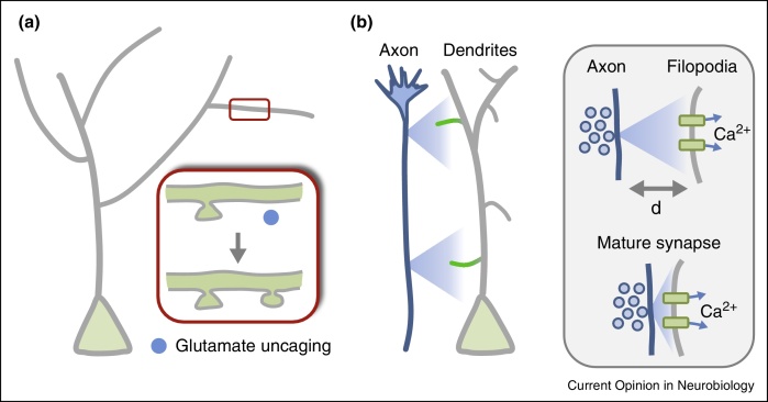
The long history of probing the role of neuronal activity in the development of nervous system circuitry has recently taken an interesting turn. Although undoubtedly activity plays a critical part in the maintenance and refinement of synaptic connections, often via competitive mechanisms, evidence is building that it also drives the process of synapse formation itself. Perhaps predictably, this turns out not to be a uniform process. It seems that different circuits, indeed specific synaptic connections, are differentially sensitive to the effects of activity. We examine possible ways in which neurotransmitter may drive synapse formation, and speculate on how the environment of the developing brain may allow a different spatiotemporal range for neuronal activity to operate in the generation of connectivity.


Figures

Figure 1
Figure 1
Synaptic activity drives the formation of synaptic contacts in the developing retina. (a) Schematic diagram of the retina showing photoreceptors (blue), bipolar cells (pink) and ganglion cells (green). Synapses between bipolar and ganglion cells occur in different layers, broadly divided into ON and OFF layers. (b) Synaptic contacts (yellow) formed between two different types of ON bipolar cells (B6 and B7, pink) and a specific type of ON ganglion cell (G10, green) are modulated by activity. Expressing tetanus toxin in B6 or B7 bipolar cells (middle panel) results in a specific decrease in the number of connections formed by B6 but not by B7 neurons. Conversely, increasing the activity of bipolar cells (bottom panel) causes an increase in the number of B6 synapses, but not those formed by B7 neurons.
Figure 2
Figure 2
The role of the neurotransmitter glutamate in synapse formation. (a) Glutamate can induce the formation of functional postsynaptic spines. The diagram shows a developing neuron in the cortex with a cell body (green) and apical dendrites (gray). The inset shows a zoomed in section of the dendrite (red box), which contains an existing spine (top drawing). Glutamate is uncaged locally with a laser (blue circle), resulting in the emergence of a new postsynaptic spine. Adapted from [53]. (b) Long range communication between neurons in developing circuits. Growing axons release neurotransmitter before they form any synaptic connections (indicated by graded blue signal from axons). Filopodia from neighbouring neurons may be able to sense this neurotransmitter at a distance (indicated by the graded green response in filopodia). Inset: the release of neurotransmitter, such as glutamate, from presynaptic vesicles (blue) clustered along a growing axon could activate distant dendritic receptors (green), such as NMDA receptors, resulting in possible calcium influx and plasticity (top drawing). Such a form of long-range communication, distinct from local synaptic transmission in mature synapses (bottom drawing) could provide information for circuit assembly and synapse formation. The spatial extent of this form of communication (d) may well be larger than the few nanometers of mature synapses and may help instruct postsynaptic filopodia and dendrites during the process of synaptogenesis.

References

    1. LeVay S., Wiesel T.N., Hubel D.H. The development of ocular dominance columns in normal and visually deprived monkeys. J Comp Neurol. 1980;191:1–51. - PubMed
    1. Wiesel T.N., Hubel D.H. Single-cell responses in striate cortex of kittens deprived of vision in one eye. J Neurophysiol. 1963;26:1003–1017. - PubMed
    1. Huberman A.D., Feller M.B., Chapman B. Mechanisms underlying development of visual maps and receptive fields. Annu Rev Neurosci. 2008;31:479–509. - PMC - PubMed
    1. Katz L.C., Shatz C.J. Synaptic activity and the construction of cortical circuits. Science. 1996;274:1133–1138. - PubMed
    1. Sanes J.R., Lichtman J.W. Development of the vertebrate neuromuscular junction. Annu Rev Neurosci. 1999;22:389–442. - PubMed

Substances

LinkOut - more resources