. 2014 Mar;26(3):862-75.
doi: 10.1105/tpc.113.119495. Epub 2014 Mar 14.

Systems analysis of auxin transport in the Arabidopsis root apex

Affiliations

Systems analysis of auxin transport in the Arabidopsis root apex

Leah R Band et al. Plant Cell. 2014 Mar.

Abstract

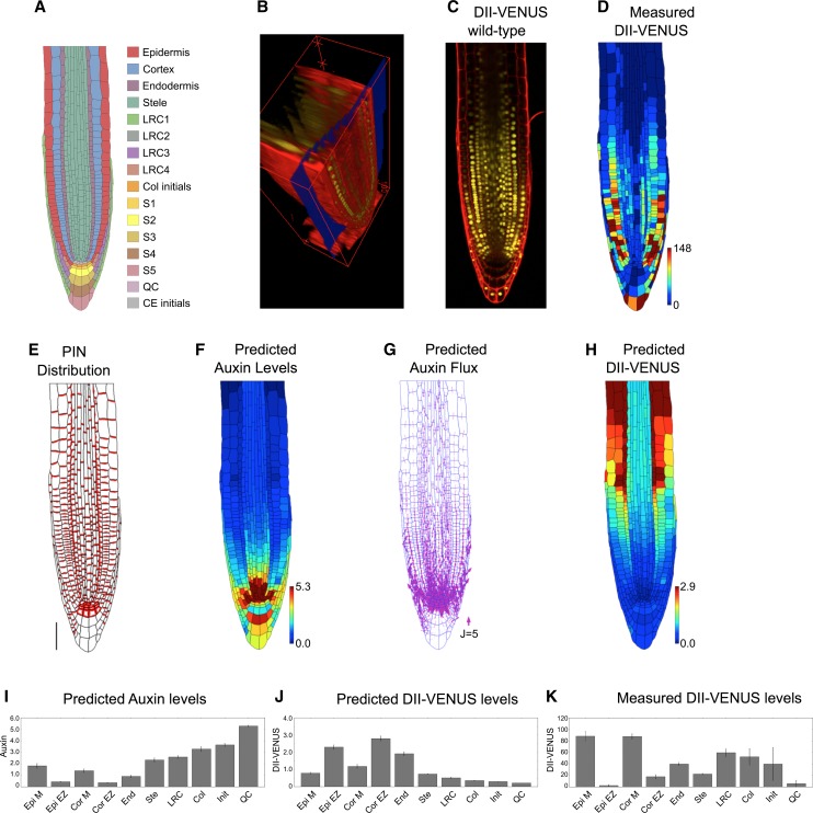
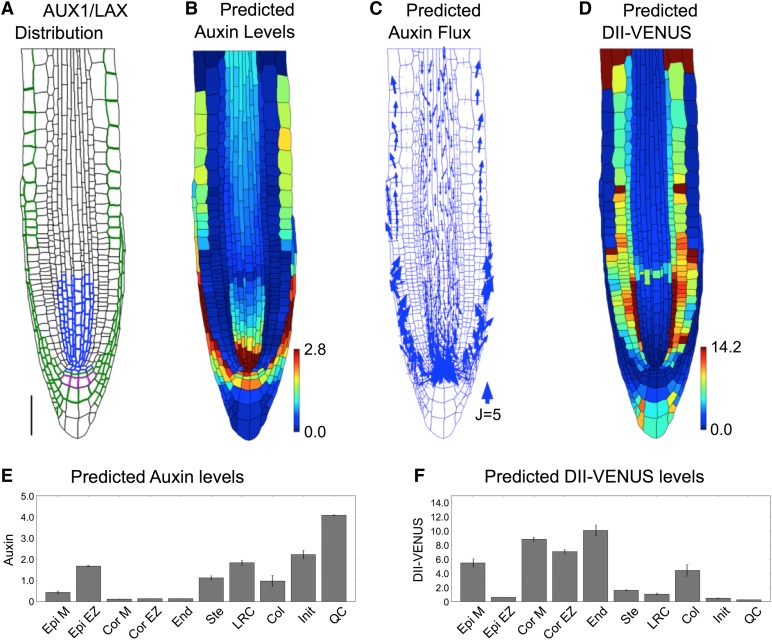
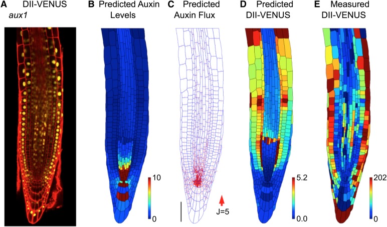
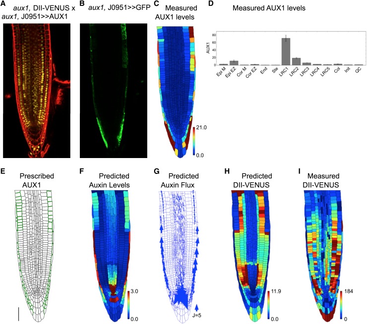
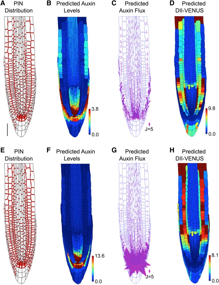
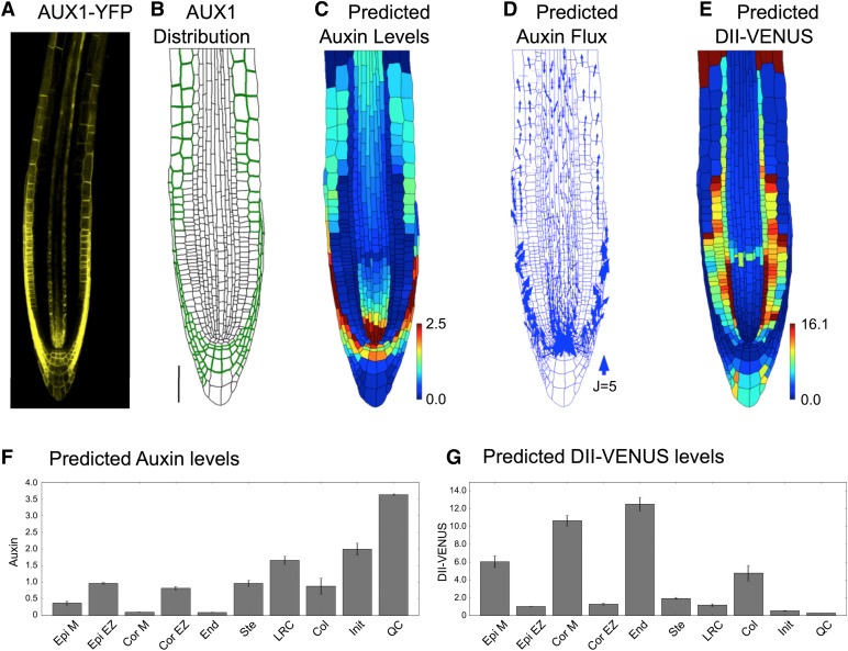
Auxin is a key regulator of plant growth and development. Within the root tip, auxin distribution plays a crucial role specifying developmental zones and coordinating tropic responses. Determining how the organ-scale auxin pattern is regulated at the cellular scale is essential to understanding how these processes are controlled. In this study, we developed an auxin transport model based on actual root cell geometries and carrier subcellular localizations. We tested model predictions using the DII-VENUS auxin sensor in conjunction with state-of-the-art segmentation tools. Our study revealed that auxin efflux carriers alone cannot create the pattern of auxin distribution at the root tip and that AUX1/LAX influx carriers are also required. We observed that AUX1 in lateral root cap (LRC) and elongating epidermal cells greatly enhance auxin's shootward flux, with this flux being predominantly through the LRC, entering the epidermal cells only as they enter the elongation zone. We conclude that the nonpolar AUX1/LAX influx carriers control which tissues have high auxin levels, whereas the polar PIN carriers control the direction of auxin transport within these tissues.


Figures

Figure 1.
Figure 1.
Auxin Transport by the PIN Efflux Carriers Alone Does Not Produce the Auxin Distribution in the Root Tip. (A) Schematic showing cell types labeled on the wild-type root geometry. CE, cortical/endodermal. (B) 3D measuring plane (blue) through a confocal stack of images produced using SurfaceProject software. (C) Output 2D interpolated slice, showing DII-VENUS (yellow) and cell geometries (red) within an Arabidopsis root tip (stained using propidium iodide). (D) Measured DII-VENUS levels extracted from the confocal image shown in (C). (E) Prescribed PIN distribution. Bar = 50 μm. (F) Predicted auxin distribution. (G) Predicted auxin fluxes (with arrow width scaling with flux). (H) Predicted DII-VENUS distribution. (I) Mean predicted auxin concentration in the different cell types. (J) Mean predicted DII-VENUS concentration in the different cell types. (K) Mean measured DII VENUS levels in the different cell types. In (D), (F), and (H), the heat map upper limit is prescribed automatically to be the 95th percentile of the predicted/measured values in each case. In (I) to (K), error bars show 1 se. Epi, epidermis; Cor, cortex; End, endodermis; Ste, stele; Col, columella; Init, columella initials; M, meristem; EZ, elongation zone. Supplemental Table 1 lists the number of cells in each category.
Figure 2.
Figure 2.
The Auxin Distribution in the Root Tip Is Due to the Interplay between PIN and AUX1/LAX-Mediated Active Transport. (A) Prescribed distributions of AUX1 (green and purple), LAX2 (blue), and LAX3 (purple). Bar = 50 μm. (B) Predicted auxin distribution. (C) Predicted auxin fluxes (with arrow width scaling with flux). (D) Predicted DII-VENUS distribution. (E) Mean predicted auxin concentration in the different cell types. (F) Mean predicted DII-VENUS concentration in the different cell types.
Figure 3.
Figure 3.
AUX1 Significantly Affects the Auxin Distribution at the Root Tip. (A) Confocal image of an aux1 null mutant root tip, reporting DII-VENUS (yellow) with propidium iodide background staining (red). (B) to (E) Predictions/measurements for the aux1 null mutant. (B) Predicted auxin distribution. (C) Predicted auxin fluxes (with arrow width scaling with flux). Bar = 50 μm. (D) Predicted DII-VENUS distribution. (E) Measured DII-VENUS levels.
Figure 4.
Figure 4.
AUX1 in LRC and Elongation Zone Epidermal Cells Is Required for the Auxin Dynamics. (A) Confocal image of aux1, DII-VENUS × aux1, J0951>>AUX1, reporting DII-VENUS (yellow) with propidium iodide background staining (red). (B) Green channel from confocal image in (A) showing J0951>>GFP (and hence AUX1), confirming AUX1 expression solely in the LRC and epidermis. (C) AUX1 fluorescence level for each cell extracted from confocal image in (B); heat map shows the average fluorescence calculated as the total fluorescence in the cell divided by the length of the cell's wall. (D) Mean AUX1 fluorescence in the different cell types. (E) Prescribed AUX1 distribution reflecting aux1, J0951>>AUX1 pattern ([C] and [D]). Bar = 50 µm. (F) Predicted auxin distribution. (G) Predicted auxin fluxes (with arrow width scaling with flux). (H) Predicted DII-VENUS distribution. (I) Measured DII-VENUS levels extracted from the confocal image in (A).
Figure 5.
Figure 5.
Effect of Periclinal PINs on Predicted Wild-Type Distributions and Fluxes with Auxin Transport Mediated by Both Influx and Efflux Carriers. (A) to (D) With inner periclinal PINs on epidermal cell membranes. Bar = 50 μm. (E) to (H) With inner periclinal PINs on epidermal, cortical, and LRC cell membranes. (A) and (E) Prescribed PIN distribution. (B) and (F) Predicted auxin distribution. (C) and (G) Predicted auxin flux (with arrow width scaling with flux). (D) and (H) Predicted DII-VENUS distribution.
Figure 6.
Figure 6.
AUX1 Expression within Elongating Cortical Cells Significantly Modifies the Auxin Distribution. (A) Localization of AUX1 using AUX1-Ypet line reveals AUX1 in the cortex of wild-type Arabidopsis root tips. (B) Prescribed AUX1 positions based on the AUX1-Ypet image (shown in [A]). Bar = 50 μm. (C) to (G) Predictions for a wild-type root tip, using the AUX1 distribution shown in (B). (C) Predicted auxin distribution. (D) Predicted auxin fluxes (with arrow width scaling with flux). (E) Predicted DII-VENUS distribution. (F) Mean predicted auxin concentration in the different cell types. (G) Mean predicted DII-VENUS concentration in the different cell types.
Figure 7.
Figure 7.
The SimuPlant GUI Consists of Five Main Components. (A) Model specification, which allows different cell geometries to be selected, model parameters to be altered, and carrier classes to be included or excluded from cell files. (B) Simulation visualization, showing the output for the modeled species in the cell geometry for all time steps. (C) Simulation Control, which allows the simulation on the server to be started or stopped and to control the number and duration of modeled time steps. (D) Simulation cell output, which shows the model output for any cell selected in the simulation visualization (indicated by green dashed lines around the cell wall). (E) Task window, which lists progress of tasks sent to and received from the server.

References

    1. Abas L., Benjamins R., Malenica N., Paciorek T., Wiśniewska J., Moulinier-Anzola J.C., Sieberer T., Friml J., Luschnig C. (2006). Intracellular trafficking and proteolysis of the Arabidopsis auxin-efflux facilitator PIN2 are involved in root gravitropism. Nat. Cell Biol. 8: 249–256. Erratum. Nat. Cell Biol. 8: 424. - PubMed
    1. Band L.R., King J.R. (2012). Multiscale modelling of auxin transport in the plant-root elongation zone. J. Math. Biol. 65: 743–785. - PubMed
    1. Band L.R., et al. (2012). Root gravitropism is regulated by a transient lateral auxin gradient controlled by a tipping-point mechanism. Proc. Natl. Acad. Sci. USA 109: 4668–4673. - PMC - PubMed
    1. Benjamins R., Scheres B. (2008). Auxin: The looping star in plant development. Annu. Rev. Plant Biol. 59: 443–465. - PubMed
    1. Benková E., Michniewicz M., Sauer M., Teichmann T., Seifertová D., Jürgens G., Friml J. (2003). Local, efflux-dependent auxin gradients as a common module for plant organ formation. Cell 115: 591–602. - PubMed

Publication types

MeSH terms

Substances