Review
. 2014 Apr 24;123(17):2605-13.
doi: 10.1182/blood-2013-09-526277. Epub 2014 Mar 14.

Multiple roles of the coagulation protease cascade during virus infection

Affiliations
Review

Multiple roles of the coagulation protease cascade during virus infection

Silvio Antoniak et al. Blood. .

Abstract

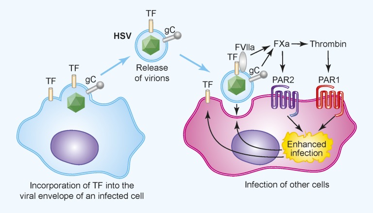
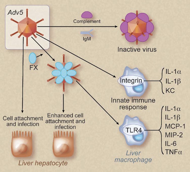
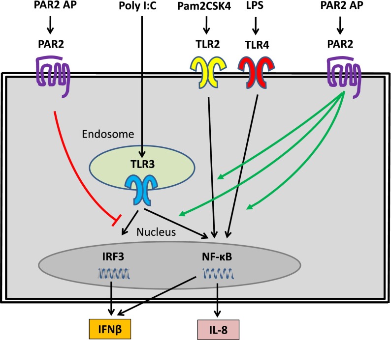
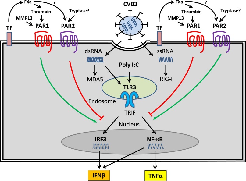
The coagulation cascade is activated during viral infections. This response may be part of the host defense system to limit spread of the pathogen. However, excessive activation of the coagulation cascade can be deleterious. In fact, inhibition of the tissue factor/factor VIIa complex reduced mortality in a monkey model of Ebola hemorrhagic fever. Other studies showed that incorporation of tissue factor into the envelope of herpes simplex virus increases infection of endothelial cells and mice. Furthermore, binding of factor X to adenovirus serotype 5 enhances infection of hepatocytes but also increases the activation of the innate immune response to the virus. Coagulation proteases activate protease-activated receptors (PARs). Interestingly, we and others found that PAR1 and PAR2 modulate the immune response to viral infection. For instance, PAR1 positively regulates TLR3-dependent expression of the antiviral protein interferon β, whereas PAR2 negatively regulates expression during coxsackievirus group B infection. These studies indicate that the coagulation cascade plays multiple roles during viral infections.


Figures

Figure 1
Figure 1
The coagulation cascade and PAR1 and PAR2 enhance HSV infection of endothelial cells. TF is incorporated into the envelope of HSV after infection of TF+ cells. Upon binding to a target cell, TF in the envelope activates the coagulation cascade. In addition, the virally encoded glycoprotein C enhances activation of FX. The local generation of FXa and thrombin activate PAR1 and PAR2 on the cells, which increases infection, possibly by modifying receptors that mediate virus binding and entry. Professional illustration by Paulette Dennis.
Figure 2
Figure 2
Binding of FX to Adv5 increases infection of hepatocytes and enhances activation of macrophages. Adv5 without FX bound is recognized by immunoglobulin M (IgM) and complement and inactivated. This form of virus has a low infectivity and mildly activates the innate immune system by binding to integrins on liver macrophages. Binding of FX to Adv5 enhances infection of liver hepatocytes by increasing the binding of virus to the cells. In addition, Adv5 coated with FX induces a more robust innate immune response in macrophages by activating the TLR4-NF-κB pathway. KC, CXCL1. Professional illustration by Debra T. Dartez.
Figure 3
Figure 3
PAR2 modulates TLR signaling in human epithelial cell lines. PAR2 positively regulated TLR2, TLR3, and TLR4 activation of NF-κB and IL-8 expression but negatively regulated TLR3 activation of IRF3 and IFN-β expression. The TLR-specific ligands are shown together with PAR2 agonist peptide (AP), which was used to activate PAR2.
Figure 4
Figure 4
PAR1 and PAR2 modulate TLR3-dependent expression of IFN-β in CFs. CVB3 infection of CFs leads to the release of ssRNA and the replication intermediate dsRNA into the cytosol. These foreign nucleic acids are recognized by TLR3 in endosomes and the cytoplasmic PRRs retinoic acid–inducible gene I (RIG-I) and melanoma differentiated gene 5 (MDA5). The TLR3-IFN-β pathway plays a central role in the innate immune response to CVB3 infection. Activation of PAR1 by TF-dependent generation of thrombin and MMP13 enhances TLR3-dependent activation of IRF3 and induction of IFN-β but suppresses TLR3-dependent activation of NF-κB and TNF-α expression. In contrast, activation of PAR2, possibly by FXa and/or tryptase, inhibits TLR3-dependent induction of IFN-β expression but enhances TLR3-dependent induction of TNF-α.

Similar articles

Cited by

References

    1. Takeuchi O, Akira S. Pattern recognition receptors and inflammation. Cell. 2010;140(6):805–820. - PubMed
    1. Beutler BA. TLRs and innate immunity. Blood. 2009;113(7):1399–1407. - PMC - PubMed
    1. Medzhitov R. Recognition of microorganisms and activation of the immune response. Nature. 2007;449(7164):819–826. - PubMed
    1. Li C, Li J, Li Y, et al. Crosstalk between platelets and the immune system: old systems with new discoveries. Adv Hematol. 2012;2012:384685. - PMC - PubMed
    1. Gorbea C, Makar KA, Pauschinger M, et al. A role for Toll-like receptor 3 variants in host susceptibility to enteroviral myocarditis and dilated cardiomyopathy. J Biol Chem. 2010;285(30):23208–23223. - PMC - PubMed

Publication types

MeSH terms