

Autoantibodies to agrin in myasthenia gravis patients
- PMID: 24632822
- PMCID: PMC3954737
- DOI: 10.1371/journal.pone.0091816
Autoantibodies to agrin in myasthenia gravis patients
Abstract
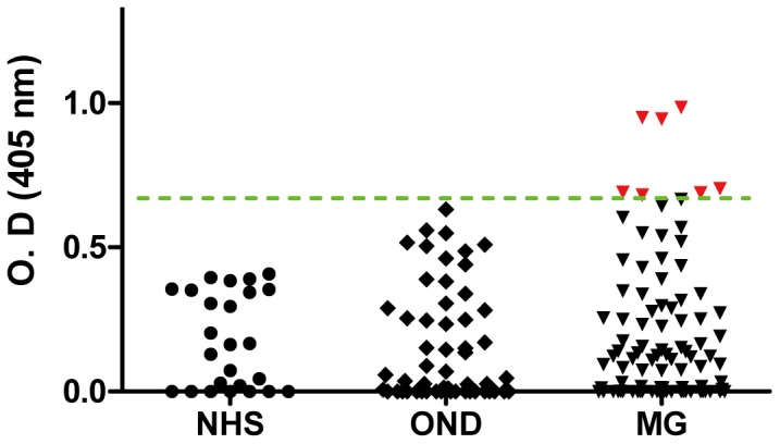
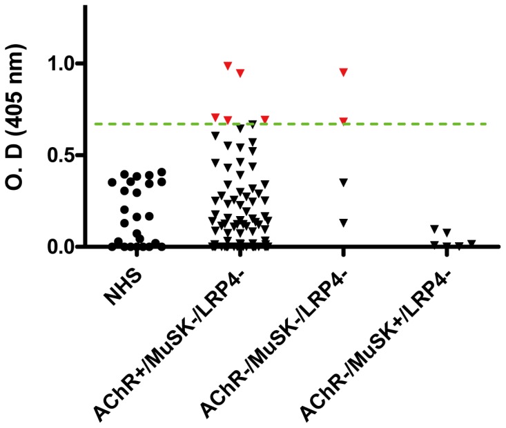
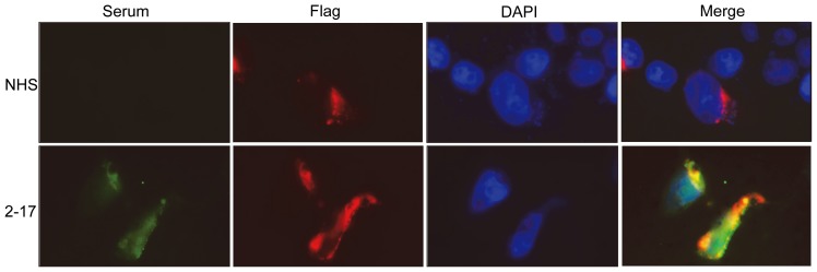
To determine if patients with myasthenia gravis (MG) have antibodies to agrin, a proteoglycan released by motor neurons and is critical for neuromuscular junction (NMJ) formation, we collected serum samples from 93 patients with MG with known status of antibodies to acetylcholine receptor (AChR), muscle specific kinase (MuSK) and lipoprotein-related 4 (LRP4) and samples from control subjects (healthy individuals and individuals with other diseases). Sera were assayed for antibodies to agrin. We found antibodies to agrin in 7 serum samples of MG patients. None of the 25 healthy controls and none of the 55 control neurological patients had agrin antibodies. Two of the four triple negative MG patients (i.e., no detectable AChR, MuSK or LRP4 antibodies, AChR-/MuSK-/LRP4-) had antibodies against agrin. In addition, agrin antibodies were detected in 5 out of 83 AChR+/MuSK-/LRP4- patients but were not found in the 6 patients with MuSK antibodies (AChR-/MuSK+/LRP4-). Sera from MG patients with agrin antibodies were able to recognize recombinant agrin in conditioned media and in transfected HEK293 cells. These sera also inhibited the agrin-induced MuSK phosphorylation and AChR clustering in muscle cells. Together, these observations indicate that agrin is another autoantigen in patients with MG and agrin autoantibodies may be pathogenic through inhibition of agrin/LRP4/MuSK signaling at the NMJ.
Conflict of interest statement
Figures


References
-
- Vincent A, Palace J, Hilton-Jones D (2001) Myasthenia gravis. Lancet 357: 2122–2128. - PubMed
-
- Phillips LH Jr (2003) The epidemiology of myasthenia gravis. Ann N Y Acad Sci 998: 407–412. - PubMed
-
- Alshekhlee A, Miles JD, Katirji B, Preston DC, Kaminski HJ (2009) Incidence and mortality rates of myasthenia gravis and myasthenic crisis in US hospitals. Neurology 72: 1548–1554. - PubMed
-
- Richman DP, Agius MA (2003) Treatment of autoimmune myasthenia gravis. Neurology 61: 1652–1661. - PubMed
Publication types
MeSH terms
Substances
LinkOut - more resources
Full Text Sources
Other Literature Sources
Medical
Miscellaneous
