. 2014 Mar 14;9(3):e91344.
doi: 10.1371/journal.pone.0091344. eCollection 2014.

Structural studies of planctomycete Gemmata obscuriglobus support cell compartmentalisation in a bacterium

Affiliations

Structural studies of planctomycete Gemmata obscuriglobus support cell compartmentalisation in a bacterium

Evgeny Sagulenko et al. PLoS One. .

Abstract

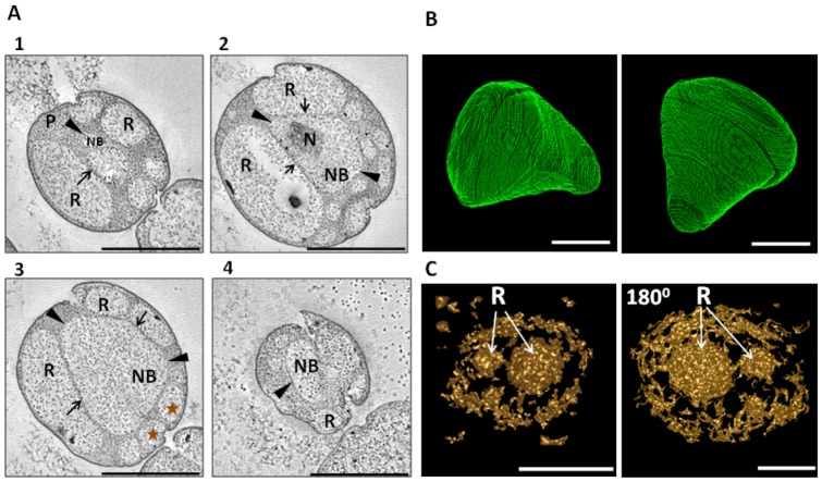
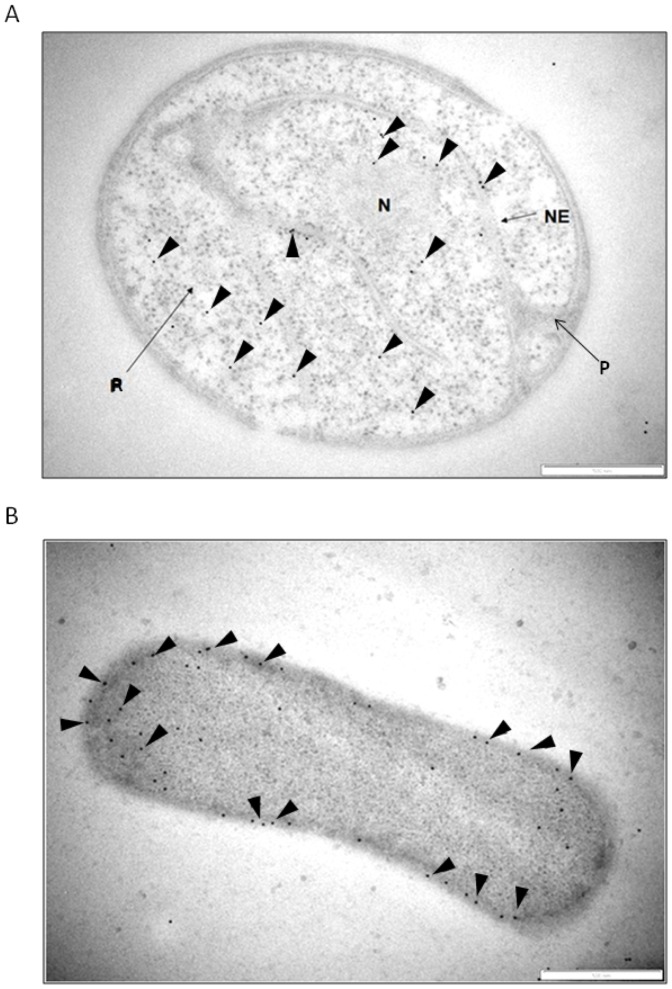
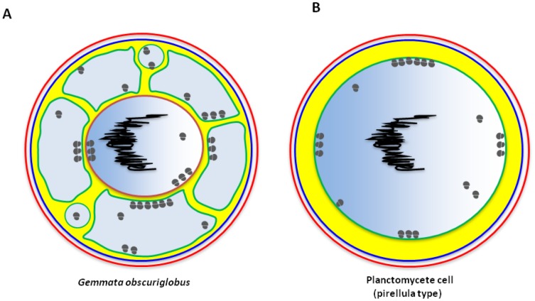
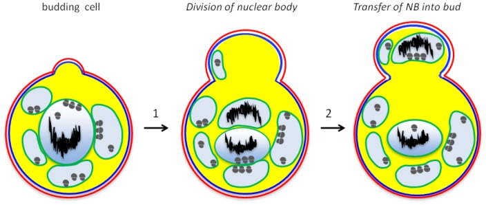
Members of phylum Planctomycetes have been proposed to possess atypical cell organisation for the Bacteria, having a structure of sectioned cells consistent with internal compartments surrounded by membranes. Here via electron tomography we confirm the presence of compartments in the planctomycete Gemmata obscuriglobus cells. Resulting 3-D models for the most prominent structures, nuclear body and riboplasm, demonstrate their entirely membrane - enclosed nature. Immunogold localization of the FtsK protein also supports the internal organisation of G.obscuriglobus cells and their unique mechanism of cell division. We discuss how these new data expand our knowledge on bacterial cell biology and suggest evolutionary consequences of the findings.


Conflict of interest statement

Competing Interests: The authors have declared that no competing interests exist.

Figures

Figure 1
Figure 1. Tomographic reconstruction of a G. obscuriglobus cell and 3D models for nuclear envelope and riboplasm.
A) Transmission electron micrographs of thick-sectioned cryosubstituted (high-pressure frozen) cells showing internal G. obscuriglobus compartments. Nuclear body (NB) contains the nucleoid DNA (N), areas of riboplasm (R) contain ribosomes only and no fibrillar nucleoid DNA, and paryphoplasm (P) is ribosome-free. Areas where nuclear body envelope is surrounded by a single membrane are indicated by arrowheads, and where areas of this envelope surrounded by a double membrane by arrows. Numbers 1-4 indicate the order of appearance of a particular image within the tilt-series. Double-membrane nuclear envelope conformation in successive tilt-series is consistent with a continuous surface of double-membrane sheet in these regions, and consistency with membrane continuity is preserved also where single membrane appears to comprise the envelope in certain regions. Stars indicate the riboplasm vesicles used for the 3D model generation (Figure 1C). The whole cell reconstruction can be viewed in Movies S1. Bar, 1 µm. (B) and (C) 3D models based on the results of electron tomography, from the cell viewed in Figure 1A (see also Movies S1), with extrapolations and manual adjustments every 10 slices. B) Nuclear body shown from two different angles. Bar marker, 500 µm. C) Riboplasm compartment (R) in the form of vesicles, completely surrounded by membranes, from front and back side (180°) views. Bar markers, 500 µm and 200 µm for the back-side view figure.
Figure 2
Figure 2. Distribution of the FtsK protein in G. obscuriglobus (A) and E.coli (B) cells.
Immunogold labelling was performed on high-pressure frozen, cryosubstituted, and then thin-sectioned cells. A) In G. obscuriglobus cells FtsK is localised mostly to the interior of nuclear body (NB) and in riboplasm (R) compartments, but not to paryphoplasm (P). B) Instead, in E.coli cells FtsK is distributed along the cell periphery (arrowheads). Bar marker, 500 nm. Arrowheads indicate gold particles. Bar marker, 500 nm.
Figure 3
Figure 3. Proposed cell organisations of planctomycetes.
A) The cell plan for G.obscuriglobus proposed in the current publication. This cell plan mostly follows the established view in past publications (as in [4]) including ribosome-less paryphoplasm and intracytoplasmic membrane (ICM), except for presentation of riboplasm, which now appears as multiple vesicles surrounding the nuclear body. Cell wall is indicated in red; cytoplasmic membrane - in dark blue; paryphoplasm - in yellow; riboplasm and nuclear body interior - in light blue; intracytoplasmic membrane - in green; inner nuclear body membrane - in brown; nucleoid DNA - in black; ribosomes – grey circles. B) The cell plan for Pirellula, which is considered as “simplest” among planctomycetes. A major internal compartment defined by an intracytoplasmic membrane (internal to cytoplasmic membrane bounding the protoplast) encloses a naked nucleoid. Unlike G. obscuriglobus, this bacterium thus does not contain a membrane-bounded nuclear body within the major internal pirellulosome compartment. Designation of the structures the same as for (A).
Figure 4
Figure 4. A model for mechanism of cell division of G. obscuriglobus cells.
Step 1, the bud appears as a hump on the surface of a cell (Figures S5 and S6 in File S1). The nuclear body is divided, before or during the formation of a bud, forming two fully enveloped structures, as shown in step 2. Finally, one of the nuclear bodies migrates into a newly formed cell (step 3). Other riboplasm vesicles not containing nucleoid DNA are also transferred into the newly formed cell (Figure S6B in File S1). Cell wall is indicated in red; plasma membrane – blue; ICM – green; paryphoplasm – yellow; riboplasm – light blue; nucleoid – black; ribosomes – grey circles.

References

    1. Lindsay MR, Webb RI, Strous M, Jetten MS, Butler MK, et al. (2001) Cell compartmentalisation in planctomycetes: novel types of structural organisation for the bacterial cell. Arch Microbiol 175: 413–429. - PubMed
    1. Jogler C, Glockner FO, Kolter R (2011) Characterization of Planctomyces limnophilus and development of genetic tools for its manipulation establish it as a model species for the phylum Planctomycetes . Appl Environ Microbiol 77: 5826–5829. - PMC - PubMed
    1. Fuerst JA, Webb RI (1991) Membrane-bounded nucleoid in the eubacterium Gemmata obscuriglobus . Proc Natl Acad Sci U S A 88: 8184–8188. - PMC - PubMed
    1. Fuerst JA, Sagulenko E (2011) Beyond the bacterium: planctomycetes challenge our concepts of microbial structure and function. Nat Rev Microbiol 9: 403–413. - PubMed
    1. Fuerst JA (2005) Intracellular compartmentation in planctomycetes. Annu Rev Microbiol 59: 299–328. - PubMed

Publication types

MeSH terms

Substances

LinkOut - more resources