. 2014 Apr;46(4):376-379.
doi: 10.1038/ng.2921. Epub 2014 Mar 16.

Recurrent PTPRB and PLCG1 mutations in angiosarcoma

Affiliations

Recurrent PTPRB and PLCG1 mutations in angiosarcoma

Sam Behjati et al. Nat Genet. 2014 Apr.

Abstract

Angiosarcoma is an aggressive malignancy that arises spontaneously or secondarily to ionizing radiation or chronic lymphoedema. Previous work has identified aberrant angiogenesis, including occasional somatic mutations in angiogenesis signaling genes, as a key driver of angiosarcoma. Here we employed whole-genome, whole-exome and targeted sequencing to study the somatic changes underpinning primary and secondary angiosarcoma. We identified recurrent mutations in two genes, PTPRB and PLCG1, which are intimately linked to angiogenesis. The endothelial phosphatase PTPRB, a negative regulator of vascular growth factor tyrosine kinases, harbored predominantly truncating mutations in 10 of 39 tumors (26%). PLCG1, a signal transducer of tyrosine kinases, encoded a recurrent, likely activating p.Arg707Gln missense variant in 3 of 34 cases (9%). Overall, 15 of 39 tumors (38%) harbored at least one driver mutation in angiogenesis signaling genes. Our findings inform and reinforce current therapeutic efforts to target angiogenesis signaling in angiosarcoma.


Figures

Figure 1
Figure 1
Distribution of mutations in PTPRB. Each circle / square / triangle represents a mutation. Red: truncating mutations. Blue: missense.
Figure 2
Figure 2
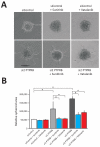
Sensitivity of PTPRB-driven angiogenesis to VEGF inhibition. A) HUVEC spheroids embedded in a fibrin gel were photographed after 24 hours of treatment (×10 magnification). B) Quantification of spheroid sprouting area. Error bars represent 1 × standard deviation. *p<0.0001.
Figure 3
Figure 3
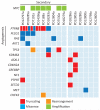
Driver variants in angiosarcoma Likely driver variants are indicated by coloured rectangles. Truncating variants (red) include nonsense, essential splice and frameshift indels. Missense substitutions are indicated in blue, amplifications in green and rearrangements in orange. Secondary angiosarcomas are either clinically classified as secondary or unclassified cases with MYC amplification.

References

    1. Young RJ, Brown NJ, Reed MW, Hughes D, Woll PJ. Angiosarcoma. Lancet Oncol. 2010;11:983–91. - PubMed
    1. Fachinger G, Deutsch U, Risau W. Functional interaction of vascular endothelial-protein-tyrosine phosphatase with the angiopoietin receptor Tie-2. Oncogene. 1999;18:5948–53. - PubMed
    1. Winderlich M, et al. VE-PTP controls blood vessel development by balancing Tie-2 activity. J Cell Biol. 2009;185:657–71. - PMC - PubMed
    1. Bamford S, et al. The COSMIC (Catalogue of Somatic Mutations in Cancer) database and website. Br J Cancer. 2004;91:355–8. - PMC - PubMed
    1. Guo T, et al. Consistent MYC and FLT4 gene amplification in radiation-induced angiosarcoma but not in other radiation-associated atypical vascular lesions. Genes Chromosomes Cancer. 2011;50:25–33. - PMC - PubMed

Publication types

MeSH terms

Substances