Review
. 2014 Aug;71(16):3059-68.
doi: 10.1007/s00018-014-1600-9. Epub 2014 Mar 17.

T cell receptor bias for MHC: co-evolution or co-receptors?

Affiliations
Review

T cell receptor bias for MHC: co-evolution or co-receptors?

Sneha Rangarajan et al. Cell Mol Life Sci. 2014 Aug.

Abstract

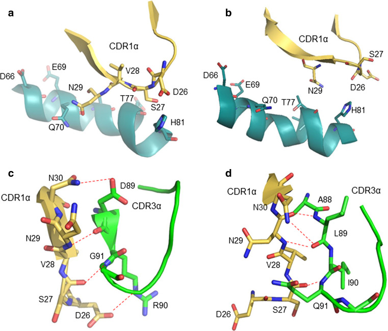
In contrast to antibodies, which recognize antigens in native form, αβ T cell receptors (TCRs) only recognize antigens as peptide fragments bound to MHC molecules, a feature known as MHC restriction. The mechanism by which MHC restriction is imposed on the TCR repertoire is an unsolved problem that has generated considerable debate. Two principal models have been advanced to explain TCR bias for MHC. According to the germline model, MHC restriction is intrinsic to TCR structure because TCR and MHC molecules have co-evolved to conserve germline-encoded TCR sequences with the ability to bind MHC, while eliminating TCR sequences lacking MHC reactivity. According to the selection model, MHC restriction is not intrinsic to TCR structure, but is imposed by the CD4 and CD8 co-receptors that promote signaling by delivering the Src tyrosine kinase Lck to TCR-MHC complexes through co-receptor binding to MHC during positive selection. Here, we review the evidence for and against each model and conclude that both contribute to determining TCR specificity, although their relative contributions remain to be defined. Thus, TCR bias for MHC reflects not only germline-encoded TCR-MHC interactions but also the requirement to form a ternary complex with the CD4 or CD8 co-receptor that is geometrically competent to deliver a maturation signal to double-positive thymocytes during T cell selection.


Figures

Fig. 1
Fig. 1
Canonical docking orientation of TCR on pMHC. a Structure of a representative TCR–pMHC complex, viewed down the MHC peptide-binding groove. The complex involves TCR 2C bound to the MHC class I molecule H-2Kb and peptide dEV8 (PDB accession code 2CKB). b Footprint of the Vα and Vβ CDR loops of TCR 2C on pMHC, illustrating the canonical diagonal docking topology. c Close superposition of the contacts of Vβ CDR1 and CDR2 with the α1 helix of I-A MHC class II in six different TCR–pMHC complexes (1U3H, 3C61, 2Z31, 2PXY, 1D9K and 3C60). All six TCRs express Vβ8.2 but different Vαs. Inset germline-encoded interactions of TCR residues CDR1β Asn31, CDR2β Tyr48, and CDR2β Glu54 with I-A residues Gln61α, Gln57α, and Lys39α
Fig. 2
Fig. 2
Editing of germline-encoded TCR–pMHC interactions by CDR3. a Interactions between CDR1α of TCR G4 and the HLA-DR1 β1 α-helix in the G4–mutTPI–DR1 complex (4E41). The side chains of contacting residues are shown. b Interactions between CDR1α of TCR E8 and the HLA-DR1 β1 α-helix in the E8–mutTPI–DR1 complex (2IAM). TCRs G4 and E8 use the same Vα region (AV13.1) but have different CDR3α sequences. CDR1α adopts different conformations in the two TCRs, resulting in different contacts with HLA-DR1. c Effect of CDR3α on the conformation of CDR1α of TCR G4. Hydrogen bonds are indicated by red dotted lines. d Effect of CDR3α on the conformation of CDR1α of TCR E8
Fig. 3
Fig. 3
TCR–pMHC complexes with atypical docking topologies. a Top view of the complex between TCR Ob.1A12 and MBP bound to HLA-DR2a (1FYT). The footprint of Ob.1A12 on MPB–DR2a is shifted towards the N-terminus of the bound peptide compared to the canonical docking mode (Fig. 1b), and most contacts to MHC are mediated by the CDR3 loops. b Side view of the complex between TCR Hy.1B11 and MBP bound to HLA-DQ (3PL6). The highly tilted binding mode of TCR Hy.1B11 prevents the Vα domain from contacting MHC. c Side view of the complex between TCR CA5 and a bulged viral peptide bound to HLA-B35 (4JRX). The TCR straddles the central region of the bound peptide but makes limited contacts with MHC. d Structure of the complex between TCR SB47 and a bulged viral peptide bound to HLA-B35 (4JRY). The TCR largely circumvents the bulged peptide by establishing an extensive footprint on the extreme N-terminal end of the MHC molecule
Fig. 4
Fig. 4
Orientation of TCR and CD4 in TCR–pMHC–CD4 complexes. a Crystal structure of a TCR–pMHC–CD4 complex (MS2-3C8–MBP–DR4–CD4) oriented as if the TCR and CD4 molecules are attached to the T cell at the bottom and the MHC class II molecules is attached to an opposing APC at the top (3T0E) [14]. b Top view of the MS2-3C8–MBP–DR4–CD4 complex, as if looking down on the T cell. The membrane-proximal TCR Cα/Cβ domains and the CD4 D4 domain are depicted in surface representation. Other domains and pMHC are omitted for clarity. The proposed arrangement of the ectodomains of CD3εγ and CD3εδ [36] is shown in relation to docking sites identified by mutational analyses [–36]. The Ig-like ectodomains of CD3εγ and CD3εδ are drawn as orange ovals. In this arrangement, only CD3γ and CD3δ contact the TCR. CD3ε projects away from the TCR, towards CD4. c Bottom view of the MS2-3C8–MBP–DR4–CD4 complex, as if looking up from inside the T cell. On the left side, the C-termini of the extracellular portions of the α and β chains of TCR MS2-3C8, as defined in the crystal structure [14], are indicated by pink and blue spheres, respectively. On the right side, the C-terminus of the extracellular portion of CD4 in the complex with MS2-3C8 and HLA-DR4 is marked by an orange sphere labeled MS2-3C8. The right side also shows the predicted position of the C-terminus of CD4 in 15 hypothetical ternary complexes constructed using other TCR–pMHC class II structures [human: HA1.7 (1JH8), Ob.1A12 (1YMM), 3A6 (1ZGL), E8 (2IAM), Hy.1B11 (3PL6), G4 (4E41); SP3.4 (4GG6); Ani2.3 (4H1L); mouse: B3K506 (3C5Z), 2W20 (3C6L), YAe62 (3C60), 21.30 (3MBE); J806.B5 (3RDT); 2B4 (3QIB); 226 (3QIU)]. In each case, the C-terminus of CD4 is marked by a black sphere labeled with the name of the corresponding TCR. The TCR–pMHC–CD4 complexes were modeled by superposing each TCR–pMHC class II structure onto the MS2-3C8–MBP–DR4–CD4 complex through the Cα/Cβ domains of the TCRs. With the exception of TCR Ob.1A12, the C-termini of CD4 in the modeled TCR–pMHC–CD4 complexes form a cluster that includes the C-terminus of CD4 in the MS2-3C8–MBP–DR4–CD4 complex. Variability in the position of the CD4 membrane anchor point is due to differences in the docking orientation of the TCR–pMHC complexes

References

    1. Tonegawa S. Somatic generation of antibody diversity. Nature. 1983;302:575–581. doi: 10.1038/302575a0. - DOI - PubMed
    1. Marrack P, Scott-Browne JP, Dai S, Gapin L, Kappler JW. Evolutionarily conserved amino acids that control TCR–MHC interaction. Annu Rev Immunol. 2008;26:171–203. doi: 10.1146/annurev.immunol.26.021607.090421. - DOI - PMC - PubMed
    1. Feng D, Bond CJ, Ely LK, Maynard J, Garcia KC. Structural evidence for a germline-encoded T cell receptor–major histocompatibility complex interaction ‘codon’. Nat Immunol. 2007;8:975–983. doi: 10.1038/ni1502. - DOI - PubMed
    1. Scott-Browne JP, White J, Kappler JW, Gapin L, Marrack P. Germline-encoded amino acids in the αβ T-cell receptor control thymic selection. Nature. 2009;458:1043–1046. doi: 10.1038/nature07812. - DOI - PMC - PubMed
    1. Adams JJ, Narayanan S, Liu B, Birnbaum ME, Kruse AC, Bowerman NA, Chen W, Levin AM, Connolly JM, Zhu C, Kranz DM, Garcia KC. T cell receptor signaling is limited by docking geometry to peptide–major histocompatibility complex. Immunity. 2011;35:681–693. doi: 10.1016/j.immuni.2011.09.013. - DOI - PMC - PubMed

Publication types

MeSH terms

Substances