

doi: 10.1038/nmeth.2887.
Epub 2014 Mar 16.
Accurate macromolecular structures using minimal measurements from X-ray free-electron lasers
Affiliations
- PMID: 24633409
- PMCID: PMC4008696
- DOI: 10.1038/nmeth.2887
Item in Clipboard
Accurate macromolecular structures using minimal measurements from X-ray free-electron lasers
Nat Methods.
2014 May.
Erratum in
- Nat Methods. 2015 Jul;12(7):692
Abstract
X-ray free-electron laser (XFEL) sources enable the use of crystallography to solve three-dimensional macromolecular structures under native conditions and without radiation damage. Results to date, however, have been limited by the challenge of deriving accurate Bragg intensities from a heterogeneous population of microcrystals, while at the same time modeling the X-ray spectrum and detector geometry. Here we present a computational approach designed to extract meaningful high-resolution signals from fewer diffraction measurements.
Conflict of interest statement
The authors declare no competing financial interests.
Figures
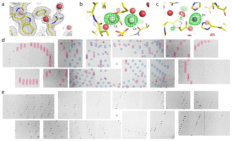
(a) 2mFo−DFc
electron density contoured at 1 σ (gray mesh) with water
molecules shown as red spheres. (b)
mFo−DFc difference
density map contoured at +3 σ (green mesh) and −3
σ (red mesh) showing binding sites for two of the four Ca ions
and (c) the single Zn ion. (d) Detail of two crystal lattices
found on the same diffraction image. Modeled spot positions assigned to the different
lattices are shown in red and blue, respectively. The sample-detector distance of 135 mm
corresponds to a resolution of 2.15 Å at the edges. (e) Detail from a
different diffraction image. Increasing radial spot elongation is observed with distance
from the beam center (blue cross).
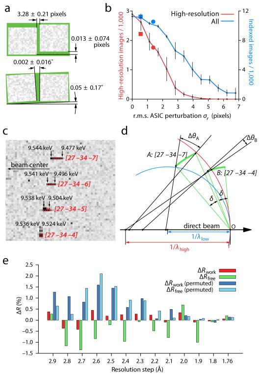
(a) Aggregate relative positions (top) and rotations (bottom) of 32 pairs of
application-specific integrated circuits (ASICs), each pair bump-bonded to a pixel array
sensor of the CSPAD detector. The two ASICs on each sensor are manufactured to be aligned
along the long axis, separated by a 3.0-pixel gap. These calibration results bear out this
expectation within the tolerances shown. (b) Impact of positional accuracy on
the indexing and integration success rate. Perturbing the ASICs away from their true
positions reduces both the total number of indexed images (blue) and the number of images
that contain successfully integrated reflections at high (1.8–2.2 Å)
resolution (red). Error bars are the standard deviation from five different sets of
perturbations drawn from a twodimensional normal distribution with a standard deviation
σr. Separate perturbations were drawn for each
ASIC. Squares: failure to apply final subpixel corrections from iterative least squares
refinement. Circles: failure to apply nearest-whole pixel corrections. (c)
Detail of four Bragg reflections on a thermolysin diffraction pattern, showing pronounced
(seven pixel) radial elongation for the [27 –34
–7] reflection and lesser elongation for those nearby. Solution
of Bragg’s law for each pixel (black arrows) identifies the spread of photon
energies that contribute to each reflection. Red disks delineate integration masks from a
three-parameter model with wavelength limits λhigh
= 1.297 (9.556 keV) and λlow = 1.313
(9.443 keV), and full-width mosaic spread δ =
0.174°. (d) Reciprocal space diagram indicating how different-shaped
reflections arise. Reciprocal lattice points (arcs) all have a constant angular extent
δ due to their mosaic spread. Points are in reflecting
condition if they are within the zone between the high-energy (red) and low-energy (blue)
Ewald spheres. Therefore, a greater fraction of the [27 –34
–7] mosaic distribution is within the reflecting condition,
leading to a reflection that subtends a greater radial angle
Δθ. (e) Paired refinements of the
thermolysin structure. Red and green bars indicate the change in
Rwork and Rfree, respectively,
as higher-resolution data are added to the refinement. Dark and light blue bars show
changes to the R-factors when the newly added high-resolution structure
factors are randomly permuted. The data are interpreted as containing statistically
significant signal for the resolution shells where
ΔRfree is continuously negative,
i.e. out to 2.1 Å.
References
-
- Neutze R, et al. Nature. 2000;406:752–757. - PubMed
Publication types
MeSH terms
Substances
Associated data
- Actions
Grants and funding
LinkOut - more resources
Full Text Sources
Other Literature Sources
