. 2014 May 15;306(10):H1426-34.
doi: 10.1152/ajpheart.01019.2013. Epub 2014 Mar 14.

IL-6 regulates adipose deposition and homeostasis in lymphedema

Affiliations

IL-6 regulates adipose deposition and homeostasis in lymphedema

Daniel A Cuzzone et al. Am J Physiol Heart Circ Physiol. .

Abstract

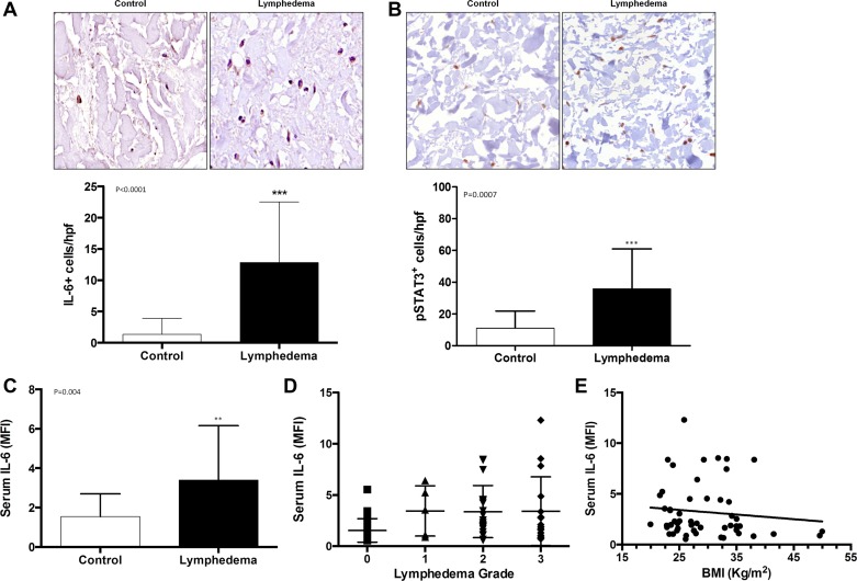
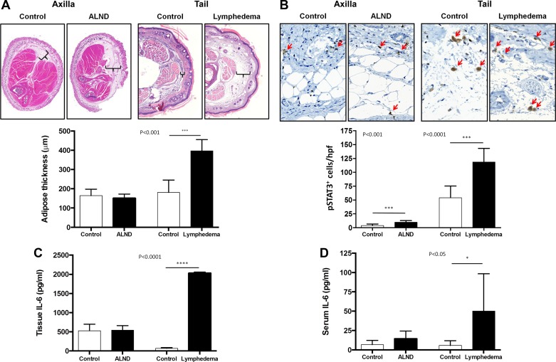
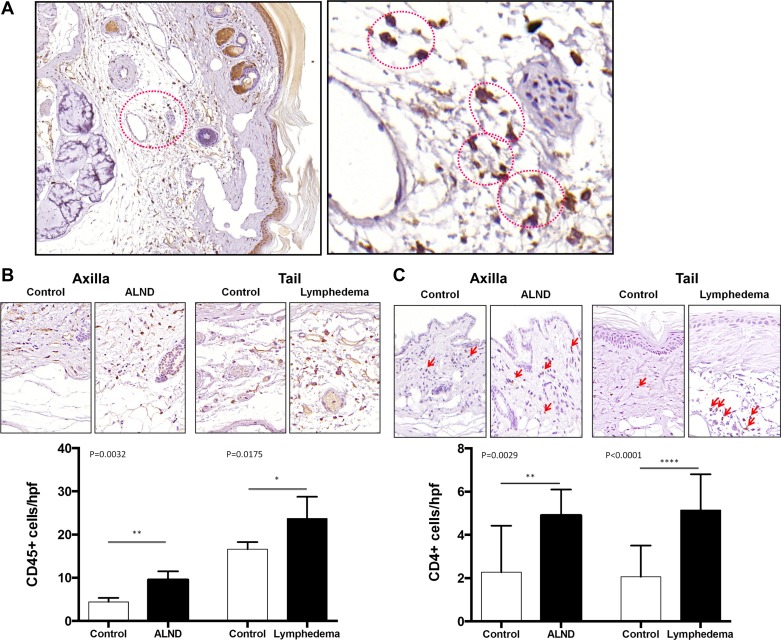
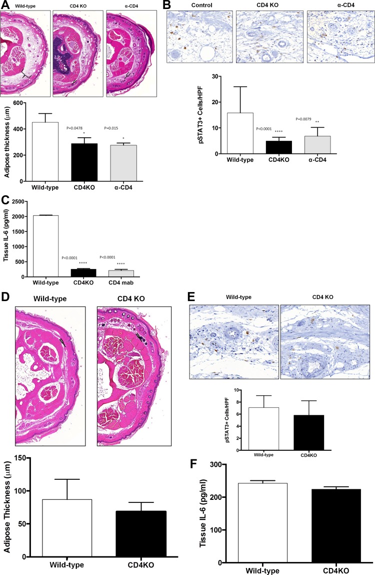
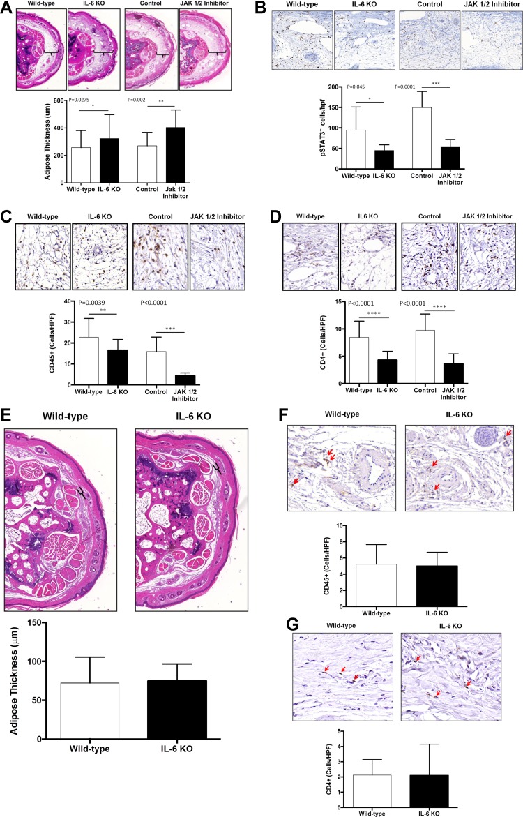
Lymphedema (LE) is a morbid disease characterized by chronic limb swelling and adipose deposition. Although it is clear that lymphatic injury is necessary for this pathology, the mechanisms that underlie lymphedema remain unknown. IL-6 is a known regulator of adipose homeostasis in obesity and has been shown to be increased in primary and secondary models of lymphedema. Therefore, the purpose of this study was to determine the role of IL-6 in adipose deposition in lymphedema. The expression of IL-6 was analyzed in clinical tissue specimens and serum from patients with or without LE, as well as in two mouse models of lymphatic injury. In addition, we analyzed IL-6 expression/adipose deposition in mice deficient in CD4(+) cells (CD4KO) or IL-6 expression (IL-6KO) or mice treated with a small molecule inhibitor of IL-6 or CD4 depleting antibodies to determine how IL-6 expression is regulated and the effect of changes in IL-6 expression on adipose deposition after lymphatic injury. Patients with LE and mice treated with lymphatic excision of the tail had significantly elevated tissue and serum expression of IL-6 and its downstream mediator. The expression of IL-6 was associated with adipose deposition and CD4(+) inflammation and was markedly decreased in CD4KO mice. Loss of IL-6 function resulted in significantly increased adipose deposition after tail lymphatic injury. Our findings suggest that IL-6 is increased as a result of adipose deposition and CD4(+) cell inflammation in lymphedema. In addition, our study suggests that IL-6 expression in lymphedema acts to limit adipose accumulation.

Keywords: IL-6; adipose; inflammation; lymphedema; serum levels.


Figures

Fig. 1.
Fig. 1.
Patients with lymphedema have increased expression of IL-6 both locally and systemically. A: representative high power (40×) photomicrographs and quantification of IL-6+cells/high-powered fields (HPF) in matched clinical lymphedema and control specimens. B: representative high power (40×) photomicrographs and quantification of pSTAT3+cells/HPF in matched clinical lymphedema and control specimens. C: quantification of serum IL-6 expression in patients with and without lymphedema [MFI (median fluorescent intensity)]. D: correlation of serum IL-6 and lymphedema grade. E: correlation of serum IL-6 and body mass index (BMI; in kg/m2).
Fig. 2.
Fig. 2.
Expression of IL-6 correlates with adipose deposition after lymphatic injury. A: representative hematoxylin and eosin (H&E) low power (2.5×) cross-sectional photomicrographs and quantification of fat thickness in the forelimb of mouse control/axillary lymph node dissection (ALND) and tail control/tail lymphedema specimens 6 wk after surgery. Brackets indicate subcutaneous adipose tissue thickness. B: representative high power (40×) photomicrographs and quantification of pSTAT3+cells/HPF in the forelimb of mouse control/ALND and tail control/lymphedema specimens. C and D: ELISA quantification of tissue IL-6 (C) and serum IL-6 (D) in both the axillary dissection model and tail lymphedema model with their respective controls.
Fig. 3.
Fig. 3.
Chronic inflammation is associated with adipose deposition and IL-6 expression. A: representative low (10×) and high power (40×) photomicrographs of crown-like structures in the adipose tissue of the murine tails following lymphatic ablation. Red circles denote the dead adipocyte and surrounding CD45+ cells. B: representative high power (40×) photomicrographs and quantification of CD45+ cells/HPF in the forelimb of mouse control/ALND and tail control/lymphedema specimens 6 wk after surgery. C: representative high power (40×) photomicrographs and quantification of CD4+ cells/HPF in the forelimb of mouse control/ALND and tail control/lymphedema specimens 6 wk after surgery.
Fig. 4.
Fig. 4.
CD4+ T lymphocyte inflammation is necessary for adipose deposition and IL-6 expression. A: representative low-power (5×) cross-sectional photomicrographs and quantification of adipose tissue thickness in tails of wild-type, CD4KO, and CD4 depleted (α-CD4) mice 6 wk after tail skin/lymphatic excision. Brackets indicate subcutaneous adipose tissue thickness. B: representative high power (40×) cross-sectional photomicrographs and quantification of pSTAT3+cells/HPF in tails of wild-type, CD4KO, and α-CD4 animals 6 wk after tail skin/lymphatic excision. C: ELISA quantification of tissue IL-6 in wild-type, CD4KO, and α-CD4 tail tissues 6 wk after skin/lymphatic excision. D: representative low-power (5×) cross-sectional photomicrographs and quantification of adipose tissue thickness in tails of wild-type and CD4KO mice preoperatively. E: representative high power (40×) cross-sectional photomicrographs and quantification of pSTAT3+ cells/HPF in tails of wild-type and CD4KO mice preoperatively. F: ELISA quantification of tissue IL-6 in wild-type and CD4KO mice preoperatively.
Fig. 5.
Fig. 5.
Loss of IL-6 function results in increased adipose deposition. A: representative low power (5×) cross-sectional photomicrographs and quantification of adipose tissue thickness in tail tissues of wild-type, IL-6KO, and JAK1/2 inhibitor treated mice and their respective controls 6 wk following tail lymphatic ablation. Brackets indicate subcutaneous adipose tissue thickness. BD: representative high power (40×) cross-sectional photomicrographs and quantification of pSTAT3+cells/HFP (B), CD45+cells/HPF (C), and CD4+cells/HPF (D) in wild-type, IL-6KO, and JAK1/2 inhibitor treated mice and their respective controls 6 wk after surgery. E: representative low power (5×) cross-sectional photomicrographs and quantification of adipose tissue thickness in tail tissues of wild-type and IL-6KO mice preoperatively. F and G: representative high power (40×) cross-sectional photomicrographs and quantification of CD45+ cells/HPF (F), and CD4+ cells/HPF (G) in tails of wild-type and IL-6KO preoperatively.

References

    1. Akira D, Taga T, Kishimoto T. Interleukin-6 in biology and medicine. Adv Immunol 54: 1–78, 1993 - PubMed
    1. Aschen S, Zampell JC, Elhadad S, Weitman E, De Brot M, Mehrara BJ. Regulation of adipogenesis by lymphatic fluid stasis: part II expression of adipose differentiation genes. Plast Reconstr Surg 129: 838–847, 2012 - PMC - PubMed
    1. Avraham T, Clavin NW, Daluvoy SV, Fernandez J, Soares MA, Cordeiro AP, Mehrara BJ. Fibrosis is a key inhibitor of lymphatic regeneration. Plast Reconstr Surg 124: 438–450, 2009 - PubMed
    1. Avraham T, Daluvoy S, Zampell J, Yan A, Haviv YS, Rockson SG, Mehrara BJ. Blockade Of transforming growth factor-β1 accelerates lymphatic regeneration during wound repair. Am J Pathol 177: 3202–3214, 2010 - PMC - PubMed
    1. Avraham T, Zampell JC, Yan A, Elhadad S, Weitman ES, Rockson SG, Bromberg J, Mehrara BJ. Th2 differentiation is necessary for soft tissue fibrosis and lymphatic dysfunction resulting from lymphedema. FASEB J 27: 1114–1126, 2013 - PMC - PubMed

Publication types

MeSH terms