. 2014 Apr 25;289(17):12177-12188.
doi: 10.1074/jbc.M113.523183. Epub 2014 Mar 14.

Trimethylation of histone H3 lysine 36 by human methyltransferase PRDM9 protein

Affiliations

Trimethylation of histone H3 lysine 36 by human methyltransferase PRDM9 protein

Mohammad S Eram et al. J Biol Chem. .

Abstract

PRDM9 (PR domain-containing protein 9) is a meiosis-specific protein that trimethylates H3K4 and controls the activation of recombination hot spots. It is an essential enzyme in the progression of early meiotic prophase. Disruption of the PRDM9 gene results in sterility in mice. In human, several PRDM9 SNPs have been implicated in sterility as well. Here we report on kinetic studies of H3K4 methylation by PRDM9 in vitro indicating that PRDM9 is a highly active histone methyltransferase catalyzing mono-, di-, and trimethylation of the H3K4 mark. Screening for other potential histone marks, we identified H3K36 as a second histone residue that could also be mono-, di-, and trimethylated by PRDM9 as efficiently as H3K4. Overexpression of PRDM9 in HEK293 cells also resulted in a significant increase in trimethylated H3K36 and H3K4 further confirming our in vitro observations. Our findings indicate that PRDM9 may play critical roles through H3K36 trimethylation in cells.

Keywords: Cancer; Enzyme Inhibitors; Enzyme Kinetics; Epigenetics; Histone Methylation; Methyltransferase.


Figures

FIGURE 1.
FIGURE 1.
PRDM9 substrate specificity. Unmodified, lysine-mutated, and lysine-methylated H3-(1–25) peptides (black bars), H3-(19–33) peptides (dark gray bars), H3-(21–44) peptides (light gray bars), and H4(1–24) peptides (white bars) were tested for PRDM9 methyltransferase activity. A reaction containing the unmodified H3-(1–25) peptide, but no enzyme, was included as a control (white bar with line pattern). Data are presented as mean ± S.D. from four experiments.
FIGURE 2.
FIGURE 2.
Monitoring H3K36 methylation by PRDM9 using mass spectrometry. The methylation state of H3K36 was monitored using LC-MS-TOF as described under “Experimental Procedures.” The mass spectra show the m/z signal for the +5-charged peptides corresponding to controls: unmethylated (H3K36me0; marked as “0” in a circle on the corresponding peak) (a); monomethylated (H3K36me1; 1) (b); dimethylated (H3K36me2; 2)(c); and trimethylated (H3K36me3; 3) (d) states. Spectra for reaction mixtures incubated for 5 h at 24 °C with unmethylated (r0), monomethylated (r1), dimethylated (r2), and trimethylated (r3) H3K36 peptide substrates confirm the methylation of H3K36 by PRDM9. The methylation state of each peptide is indicated by the number of methyl groups in a circle next to each peak. Methylation of H3K36me2 substrate after 10 (eR10min) and 45 min (eR45min) indicates the gradual reduction of dimethylated substrate and production of the trimethylated H3K36.
FIGURE 3.
FIGURE 3.
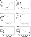
Effect of pH, ionic strength, and buffer additives on PRDM9 activity. The methyltransferase activity of PRDM9 using H3-(1–25) peptide as substrate was determined as a function of pH (a), NaCl (b), DMSO (c), Triton X-100 (d), DTT (e), and TCEP (f) as described under “Experimental Procedures.” Data points are presented as mean ± S.D. from four experiments.
FIGURE 4.
FIGURE 4.
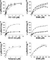
Kinetic analysis of PRDM9 methyltransferase activity. Km values were determined for H3-(1–25) peptides (H3K4me0 (▾), H3K4me1 (●), and H3K4me2 (○)) at saturation concentrations of AdoMet (SAM) (448 μm) (a) and for AdoMet with the same set of H3-(1–25) peptides (b). Similarly, Km values were determined for H3-(21–44) peptides (H3K36me0 (▾), H3K36me1 (●), and H3K36me2 (○)) at saturation concentrations of AdoMet (448 μm) (c) and for AdoMet with the same set of H3-(21–44) peptides (d). Km values were also determined for native (●) and truncated (○) H3-H4 tetramer in the presence of 350 μm AdoMet (e) and for AdoMet in the presence of 4.5 μm native (●) and 6.5 μm truncated H3-H4 tetramer (○) (f). In truncated H3-H4 tetramer, the first 11 amino acids of H3 were truncated. V, velocity. Data points are presented as mean ± S.D. from three experiments. Kinetic parameters are presented in Table 1.
FIGURE 5.
FIGURE 5.
Screening PRDM9 in 384-well format. a, the Z-factor was determined (0.82) for PRDM9 screening with H3K36me2(21–44) peptide as a substrate at concentrations of peptide and AdoMet equal to their Km values (2.5 and 62 μm, respectively). b, PRDM9 was screened against a set of known histone methyltransferase inhibitors and suramin was identified as an inhibitor with an IC50 value of 4.1 ± 0.1 μm. Experiments were performed in triplicate as described under “Experimental Procedures.”
FIGURE 6.
FIGURE 6.
H3K36 methylation on nucleosome. Reconstituted nucleosome with recombinant wild-type histone H3 (WT) and truncated histone H3 (ΔH3; missing the first 11 residues including lysine 4) were used as substrate for PRDM9, and (a) the activity was assayed by monitoring the transfer of [3H]methyl groups from [3H]AdoMet to nucleosome using the trichloroacetic acid precipitation method as described under “Experimental Procedures.” Samples of each reaction mixture (Methylated) and untreated samples (Unmethylated; control) were immunoblotted using H3K4me3 (b) and H3K36me3 (c) antibodies (upper panels for b and c). The same dot blots were immunoblotted using histone H3 antibody (IB: H3) to quantify the amount of loaded nucleosome (lower panels for b and c).
FIGURE 7.
FIGURE 7.
Assessing interactions of PRDM9 with substrates. Binding of H3-(1–25) and H3-(21–44) peptides with various H3K4 and H3K36 methylation states was monitored by ITC as described under “Experimental Procedures.” Peptide substrates and their corresponding KD values from a single run are indicated on the plots. Experiments were performed in duplicate. Mean KD values are listed in Table 1.
FIGURE 8.
FIGURE 8.
Trimethylation of H3K4 and H3K36 by PRDM9 in cells. Trimethylation of H3K36 (a) and H3K4 (b) in HEK293 cells transfected with control and PRDM9 plasmids was monitored by Western immunoblotting. c, blot quantifications indicate significant H3K4 and H3K36 trimethylation in samples from cells overexpressing PRDM9. Methylation levels of Lys-4 and Lys-36 were quantified and normalized to their respective total H3 levels. FLAG-PRDM9-transfected samples were normalized to samples transfected with FLAG Control vector. IB, immunoblot; CTL, control.
FIGURE 9.
FIGURE 9.
H3K36me2 peptide docking. The docked H3K36me2 peptide (a) recapitulates interactions observed with the backbone of the co-crystallized H3K4me2 peptide (b). H3V35 and H3P38 side chains make favorable desolvation contacts with hydrophobic clefts of the substrate-binding groove of PRDM9, and H3K37 is engaged in an electrostatic interaction with Glu-360.

References

    1. Kouzarides T. (2007) Chromatin modifications and their function. Cell 128, 693–705 - PubMed
    1. Murray K. (1964) The occurrence of ϵ-N-methyl lysine in histones. Biochemistry 3, 10–15 - PubMed
    1. Byvoet P., Shepherd G. R., Hardin J. M., Noland B. J. (1972) The distribution and turnover of labeled methyl groups in histone fractions of cultured mammalian cells. Arch. Biochem. Biophys. 148, 558–567 - PubMed
    1. Paik W. K., Kim S. (1967) ϵ-N-Dimethyllysine in histones. Biochem. Biophys. Res. Commun. 27, 479–483
    1. Hempel K., Lange H. W., Birkofer L. (1968) ϵ-N-Trimethyllysine, a new amino acid in histones. Naturwissenschaften 55, 37. - PubMed

Publication types