. 2013 Nov 1:110:79-85.
doi: 10.1016/j.electacta.2013.01.153.

The Impact of Enzyme Orientation and Electrode Topology on the Catalytic Activity of Adsorbed Redox Enzymes

Affiliations

The Impact of Enzyme Orientation and Electrode Topology on the Catalytic Activity of Adsorbed Redox Enzymes

Duncan G G McMillan et al. Electrochim Acta. .

Abstract

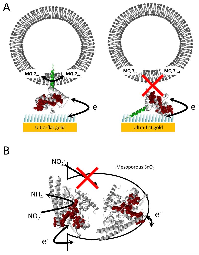
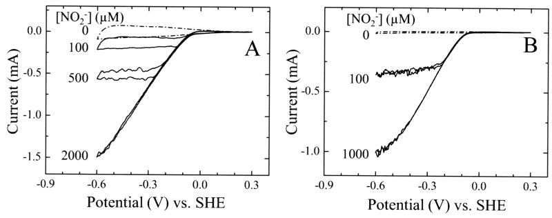
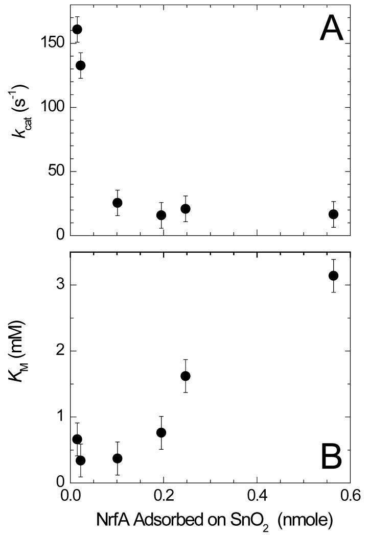
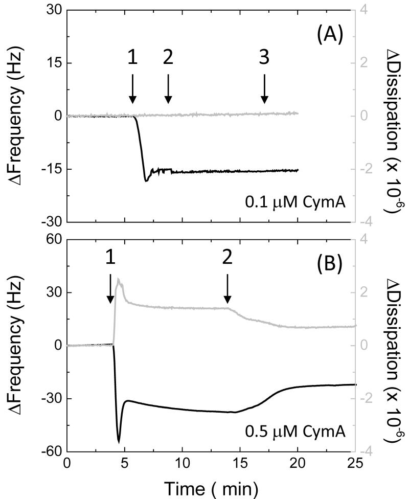
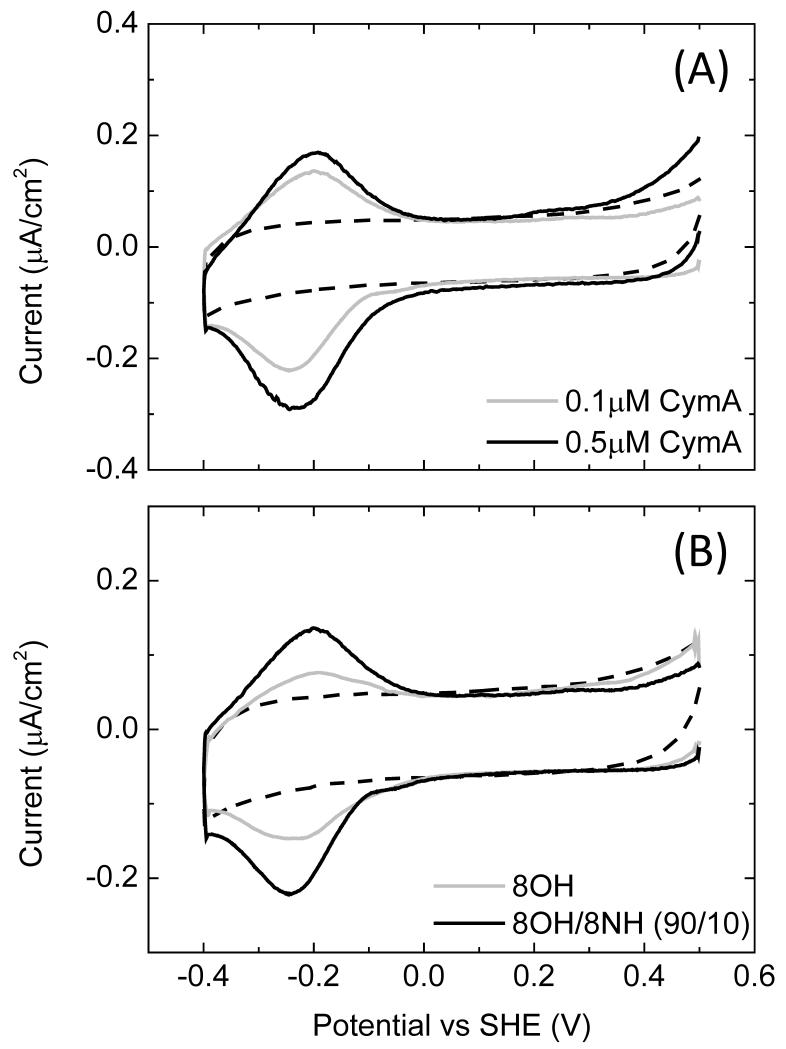
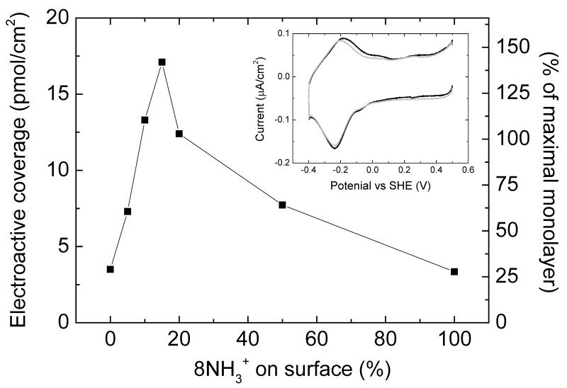
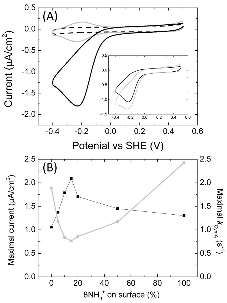
It is well established that the structural details of electrodes and their interaction with adsorbed enzyme influences the interfacial electron transfer rate. However, for nanostructured electrodes, it is likely that the structure also impacts on substrate flux near the adsorbed enzymes and thus catalytic activity. Furthermore, for enzymes converting macro-molecular substrates it is possible that the enzyme orientation determines the nature of interactions between the adsorbed enzyme and substrate and therefore catalytic rates. In essence the electrode may impede substrate access to the active site of the enzyme. We have tested these possibilities through studies of the catalytic performance of two enzymes adsorbed on topologically distinct electrode materials. Escherichia coli NrfA, a nitrite reductase, was adsorbed on mesoporous, nanocrystalline SnO2 electrodes. CymA from Shewanella oneidensis MR-1 reduces menaquinone-7 within 200 nm sized liposomes and this reaction was studied with the enzyme adsorbed on SAM modified ultra-flat gold electrodes.

Keywords: Self-assembled monolayer (SAM); cytochrome; lipid vesicle; protein-film electrochemistry (PFE); quinone.


Figures

Fig. 1
Fig. 1. Schematics of the protein film electrochemistry pursued in this work.
(A) A ribbon presentation of NrfH, a homologue of CymA, immobilised on a SAM modified gold electrode together with a lipid vesicle with MQ-7. The 8OH and 8NH3+ thiols in the SAM are shown as blue bars, the NrfH polypeptide in white (globular ‘head’ domain), green (lipophilic ‘tail’) and red (the prosthetic heme groups). Two possible orientations for the adsorption of CymA are illustrated. (B) A ribbon presentation of NrfA adsorbed within a mesoporous, nanocrystalline SnO2 electrode with the polypeptide in white and hemes in red.
Fig. 2
Fig. 2. Baseline corrected CVs of NrfA adsorbed on mesoporous, nanocrystalline SnO2 electrodes at the indicated nitrite concentrations.
Electroactive coverage of NrfA: (A) 0.56 nmol/electrode and (B) 0.02 nmol/electrode. Scan rate 30 mV/s in 50 mM Hepes, 2 mM CaCl2, pH 7.
Fig. 3
Fig. 3. Variation of kcat and KM for NrfA nitrite reduction as a function of the electroactive coverage of NrfA adsorbed on the mesoporous SnO2 electrode.
Fig. 4
Fig. 4. QCM-D results in buffer with frequency (black line, left axis) and dissipation (grey line, right axis) against time for (A) a 8OH/8NH3+ (90/10) and (B) a pure 8OH modified gold surface.
Time points: 1, (A) 0.1 and (B) 0.5 μM CymA in buffer; 2, buffer only; 3, CV between 0.5 and −0.4 V versus SHE at 10 mV/s. The plots shown are representative of triplicate experiments.
Fig. 5
Fig. 5. (A) CVs (10 mV/s) of 8OH/8NH3+ (90/10) modified gold electrode in buffer, incubated with 0.1 or 0.5 μM CymA, as indicated. (B) CVs (10 mV/s) of 8OH or 8OH/8NH3+ (90/10) modified gold electrodes, as indicated.
The CV are measured in buffer after incubation with 0.1 μM CymA. The dashed lines in (A) and (B) are CVs before incubation with CymA.
Fig. 6
Fig. 6. Electroactive coverage as determined by CV as a function of relative amount of 8NH3+ on the surface, which is given as 8NH3+/(8NH3+ + 8OH).
The surfaces were incubated with 0.5 μM CymA. (Insert) CVs (10 mV/s) of (black line) pure 8OH and (grey line) pure 8NH3+ modified gold electrode in buffer, incubated with 0.5 μM CymA.
Fig. 7
Fig. 7. (A) CVs (10 mV/s) of 8OH/8NH3+ (90/10) modified gold electrode in buffer, incubated with 0.5 μM CymA before (grey line) and after (black line) addition of POPC vesicles containing 1.0% (w/w) MQ-7. The dashed line is a CV before incubation with CymA. (Insert) idem, for (black line) pure 8OH and (grey) pure 8NH3+ modified surfaces. (B) Black line: The maximum current determined at about −0.2 V vs SHE from CVs such as shown in (A) as a function of the relative amount of 8NH3+ on the surface, which is given as 8NH3+/(8NH3+ + 8OH). Grey line: kCymA (= the activity per CymA enzyme) as a function of the relative amount of 8NH3+ on the surface.

References

    1. Leger C, Bertrand P. Chem. Rev. 2008;108:2379–2438. - PubMed
    1. Armstrong FA. Curr. Opin. Chem. Biol. 2005;9:110–117. - PubMed
    1. Zhang JD, Kuznetsov AM, Medvedev IG, Chi QJ, Albrecht T, Jensen PS, Ulstrup J. Chem. Rev. 2008;108:2737–2791. - PubMed
    1. Cracknell JA, Vincent KA, Armstrong FA. Chem. Rev. 2008;108:2439–2461. - PubMed
    1. Willner I, Yan YM, Willner B, Tel-Vered R. Fuel Cells. 2009;9:7–24.

LinkOut - more resources