. 2014 Mar 8:9:1243-60.
doi: 10.2147/IJN.S58236. eCollection 2014.

Direct role of interrod spacing in mediating cell adhesion on Sr-HA nanorod-patterned coatings

Affiliations

Direct role of interrod spacing in mediating cell adhesion on Sr-HA nanorod-patterned coatings

Jianhong Zhou et al. Int J Nanomedicine. .

Abstract

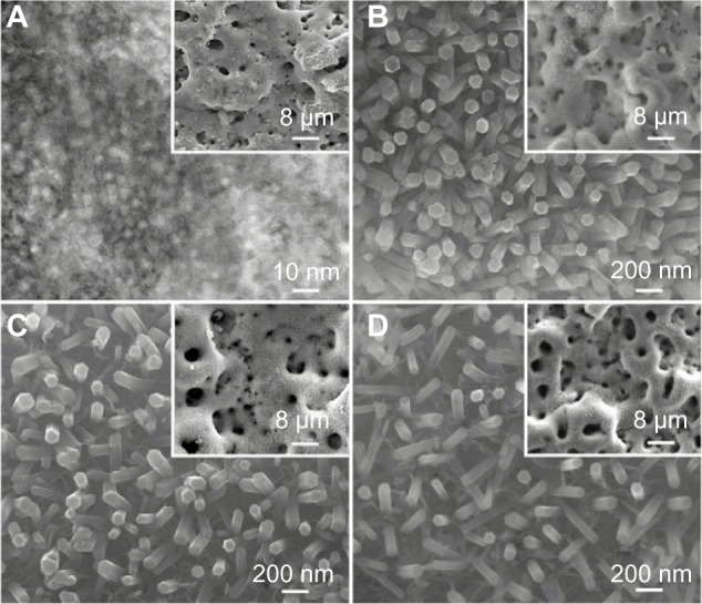
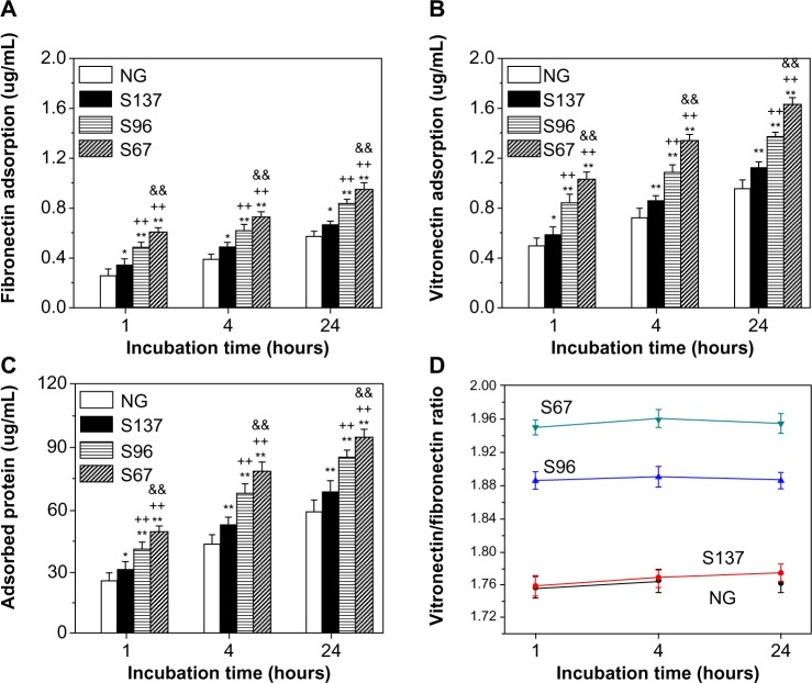
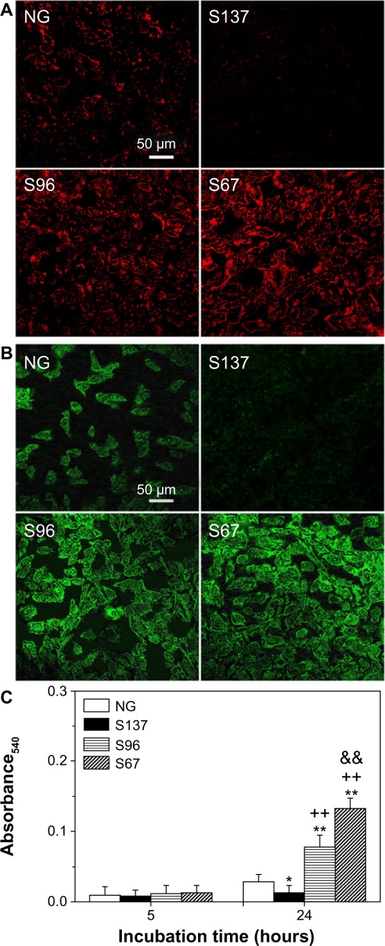
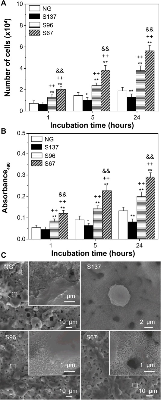
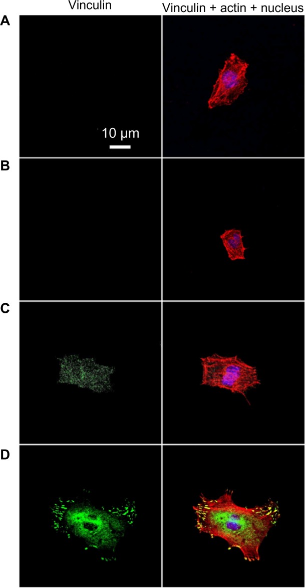
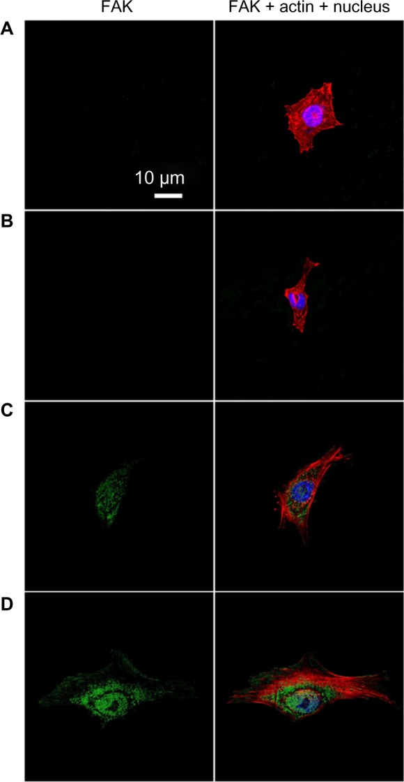
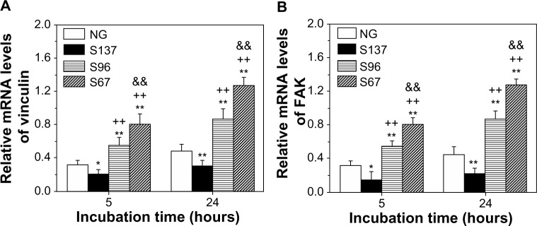
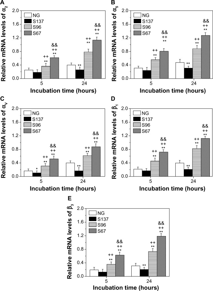
The process in which nanostructured surfaces mediate cell adhesion is not well understood, and may be indirect (via protein adsorption) or direct. We prepared Sr-doped hydroxyapatite (Sr1-HA) 3D nanorods (with interrod spacing of 67.3 ± 3.8, 95.7 ± 4.2, and 136.8 ± 8.7 nm) and 2D nanogranulate patterned coatings on titanium. Employing the coatings under the same surface chemistry and roughness, we investigated the indirect/direct role of Sr1-HA nanotopographies in the regulation of osteoblast adhesion in both serum-free and serum-containing Dulbecco's Modified Eagle/Ham's Medium. The results reveal that the number of adherent cells, cell-secreted anchoring proteins (fibronectin, vitronectin, and collagen), vinculin and focal adhesion kinase (FAK) denoted focal adhesion (FA) contact, and gene expression of vinculin, FAK, and integrin subunits (α2, α5, αv, β1, and β3), undergo significant changes in the inter-nanorod spacing and dimensionality of Sr1-HA nanotopographies in the absence of serum; they are significantly enhanced on the <96 nm spaced nanorods and more pronounced with decreasing interrod spacing. However, they are inhibited on the >96 nm spaced nanorods compared to nanogranulated 2D topography. Although the adsorption of fibronectin and vitronectin from serum are higher on 136.8 ± 8.7 nm spaced nanorod patterned topography than nanogranulated topography, cell adhesion is inhibited on the former compared to the latter in the presence of serum, further suggesting that reduced cell adhesion is independent of protein adsorption. It is clearly indicated that 3D nanotopography can directly modulate cell adhesion by regulating integrins, which subsequently mediate anchoring proteins' secretion and FA formation rather than via protein adsorption.

Keywords: anchoring protein secretion; focal adhesion; integrin; inter-nanorod spacing; nanotopography; osteoblast adhesion.


Figures

Figure 1
Figure 1
(A) TEM image taken from the surface of NG coating; SEM surface morphologies of (B) S67, (C) S96, and (D) S137 coatings; the inserts showing corresponding low magnification SEM images. Abbreviations: NG, Sr-doped hydroxyapatite nanogranule-patterned multilayer coating; SEM, scanning electron microscopy; TEM, transmission electron microscopy.
Figure 2
Figure 2
Adsorption of (A) fibronectin, (B) vitronectin, and (C) total protein as well as (D) ratio of vitronectin/fibronectin adsorbed onto the coatings after 1, 4, and 24 hours of incubation in serum-containing DMEM-12 medium. Notes: Data are presented as the mean ± SD, n=4, *P<0.05 and **P<0.01 compared with the NG coating; ++P<0.01 compared with the S137 coating; &P<0.01 compared with the S96 coating. Abbreviations: NG, Sr-doped hydroxyapatite nanogranule-patterned multilayer coating; DMEM-12, Dulbecco’s Modified Eagle/Ham’s Medium; SD, standard deviation.
Figure 3
Figure 3
Fluorescence images of (A) fibronectin and (B) vitronectin secreted by osteoblasts into ECM on NG, S137, S96, and S67 coatings after 24 hours of incubation in serum-free DMEM-12. (C) Collagen secretion in ECM by osteoblasts on the coatings after 5 and 24 hours of incubation in serum-free DMEM-12. Notes: Data are presented as the mean ± SD, n=4, *P<0.05 and **P<0.01 compared with the NG coating; ++P<0.01 compared with the S137 coating; &P<0.01 compared with the S96 coating. Abbreviations: DMEM-12, Dulbecco’s Modified Eagle/Ham’s Medium; ECM, extracellular matrix; NG, Sr-doped hydroxyapatite nanogranule-patterned multilayer coating.
Figure 4
Figure 4
(A) Counting and (B) MTT assays of osteoblasts on the NG, S137, S96, and S67 coatings after 1, 5, and 24 hours of incubation in serum-free DMEM-12. (C) Morphologies of cells on the coatings after 1 hour of incubation in serum-free DMEM-12; the inserts are corresponding magnified images. Notes: Data are presented as the mean ± SD, n=4, *P<0.05 and **P<0.01 compared with the NG coating; ++P<0.01 compared with the S137 coating; &P<0.01 compared with the S96 coating. Abbreviations: DMEM-12, Dulbecco’s Modified Eagle/Ham’s Medium; MTT, 3-(4,5-dimethylthiazol-2-yl)-2,5-diphenyltetrazolium bromide; NG, Sr-doped hydroxyapatite nanogranule-patterned multilayer coating.
Figure 5
Figure 5
Vinculin (green), actin (red), and nucleus (blue) fluorescence images of osteoblasts on the (A) NG, (B) S137, (C) S96, and (D) S67 coatings after 24 hours of incubation in serum-free DMEM-12. Abbreviations: DMEM-12, Dulbecco’s Modified Eagle/Ham’s Medium; NG, Sr-doped hydroxyapatite nanogranule-patterned multilayer coating.
Figure 6
Figure 6
FAK (green), actin (red), and nucleus (blue) fluorescence images of osteoblasts on the (A) NG, (B) S137, (C) S96, and (D) S67 coatings after 24 hours of incubation in serum-free DMEM-12. Abbreviations: DMEM-12, Dulbecco’s Modified Eagle/Ham’s Medium; FAK, focal adhesion kinase; NG, Sr-doped hydroxyapatite nanogranule-patterned multilayer coating.
Figure 7
Figure 7
(A) Vinculin and (B) FAK gene expression of osteoblasts on the coatings after 5 and 24 hours of incubation in serum-free DMEM-12. Notes: Data are presented as the mean ± SD, n=4, *P<0.05 and **P<0.01 compared with the NG coating; ++P<0.01 compared with the S137 coating; &P<0.01 compared with the S96 coating. Abbreviations: DMEM-12, Dulbecco’s Modified Eagle/Ham’s Medium; FAK, focal adhesion kinase; NG, Sr-doped hydroxyapatite nanogranule-patterned multilayer coating; mRNA, messenger ribonucleic acid.
Figure 8
Figure 8
(A) α2, (B) α5, (C) αv, (D) β1, and (E) β3 gene expression of osteoblasts on the coatings after 5 and 24 hours of incubation in serum-free DMEM-12. Notes: Data are presented as the mean ± SD, n=4, *P<0.05 and **P<0.01 compared with the NG coating; ++P<0.01 compared with the S137 coating; &P<0.01 compared with the S96 coating. Abbreviations: DMEM-12, Dulbecco’s Modified Eagle/Ham’s Medium; NG, Sr-doped hydroxyapatite nanogranule-patterned multilayer coating; mRNA, messenger ribonucleic acid; SD, standard deviation.

Similar articles

Cited by

References

    1. Anselme K. Osteoblast adhesion on biomaterials. Biomaterials. 2000;21(7):667–681. - PubMed
    1. Liu XM, Lim JY, Donahue HJ, Dhurjati R, Mastro AM, Vogler EA. Influence of substratum surface chemistry/energy and topography on the human fetal osteoblastic cell line hFOB 1.19: phenotypic and genotypic responses observed in vitro. Biomaterials. 2007;28(31):4535–4550. - PMC - PubMed
    1. Geiger B, Spatz JP, Bershadsky AD. Environmental sensing through focal adhesions. Nat Rev Mol Cell Biol. 2009;10(1):21–33. - PubMed
    1. Geiger B, Bershadsky A, Pankov R, Yamada KM. Transmembrane crosstalk between the extracellular matrix and the cytoskeleton. Nat Rev Mol Cell Biol. 2001;2(11):793–805. - PubMed
    1. Hynes RO. Integrins: bidirectional, allosteric signaling machines. Cell. 2002;110(6):673–687. - PubMed

Publication types