Review
. 2014 Mar 5:5:75.
doi: 10.3389/fphys.2014.00075. eCollection 2014.

Role of tumor associated macrophages in tumor angiogenesis and lymphangiogenesis

Affiliations
Review

Role of tumor associated macrophages in tumor angiogenesis and lymphangiogenesis

Vladimir Riabov et al. Front Physiol. .

Abstract

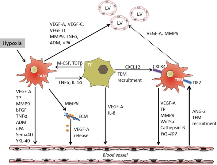
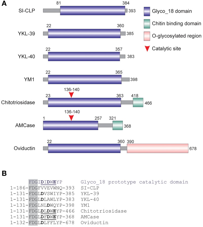
Tumor angiogenesis is an essential process for supplying rapidly growing malignant tissues with essential nutrients and oxygen. An angiogenic switch allows tumor cells to survive and grow, and provides them access to vasculature resulting in metastatic disease. Monocyte-derived macrophages recruited and reprogrammed by tumor cells serve as a major source of angiogenic factors boosting the angiogenic switch. Tumor endothelium releases angiopoietin-2 and further facilitates recruitment of TIE2 receptor expressing monocytes (TEM) into tumor sites. Tumor-associated macrophages (TAM) sense hypoxia in avascular areas of tumors, and react by production of angiogenic factors such as VEGFA. VEGFA stimulates chemotaxis of endothelial cells (EC) and macrophages. In some tumors, TAM appeared to be a major source of MMP9. Elevated expression of MMP9 by TAM mediates extracellular matrix (ECM) degradation and the release of bioactive VEGFA. Other angiogenic factors released by TAM include basic fibroblast growth factor (bFGF), thymidine phosphorylase (TP), urokinase-type plasminogen activator (uPA), and adrenomedullin (ADM). The same factors used by macrophages for the induction of angiogenesis [like vascular endothelial growth factor A (VEGF-A) and MMP9] support lymphangiogenesis. TAM can express LYVE-1, one of the established markers of lymphatic endothelium. TAM support tumor lymphangiogenesis not only by secretion of pro-lymphangiogenic factors but also by trans-differentiation into lymphatic EC. New pro-angiogenic factor YKL-40 belongs to a family of mammalian chitinase-like proteins (CLP) that act as cytokines or growth factors. Human CLP family comprises YKL-40, YKL-39, and SI-CLP. Production of all three CLP in macrophages is antagonistically regulated by cytokines. It was recently established that YKL-40 induces angiogenesis in vitro and in animal tumor models. YKL-40-neutralizing monoclonal antibody blocks tumor angiogenesis and progression. The role of YKL-39 and SI-CLP in tumor angiogenesis and lymphangiogenesis remains to be investigated.

Keywords: LYVE-1; TIE2 receptor; VEGF; chitinase-like protein; stabilin-1; tumor-associated macrophages.


Figures

Figure 1
Figure 1
Cross-talk between macrophages, tumor cells and endothelium during tumor angiogenesis and lymphangiogenesis. Tumor hypoxia and cytokine crosstalk between tumor cells (TC) and TAM results in the induction of a pro-angiogenic program in both cell types followed by the release of multiple soluble factors stimulating migration and proliferation of vascular endothelial cells (VEC) either directly or through the mobilization of extracellular matrix (ECM)-bound VEGF-A. In addition, TC and VEC are able to attract specific population of TIE-2 expressing monocytes (TEM) with high angiogenic potential. TAM and TEM-derived factors as well-induce growth of tumor lymphatic vessels (LV). Through the production of factors such as VEGF-A and MMP9 influencing growth of both lymphatic and vascular EC, TAM link processes of tumor angio- and lymphangiogenesis.
Figure 2
Figure 2
Mammalian chitinase-like proteins (CLP) belong to the family of Glyc_18-containig proteins. (A) Schematic presentation of mammalian Glyco_18 domain containing proteins. (B) Critical amino acid in catalytic sites. The characteristic FDG sequence preceding the catalytic motif is shown in the shadowed box. Catalytic amino acids are shown in bold. Complete active catalytic motifs are underlined. This research was originally published in Blood. Kzhyshkowska et al. (2006c) © the American Society of Hematology.

References

    1. Algars A., Irjala H., Vaittinen S., Huhtinen H., Sundstrom J., Salmi M., et al. (2012). Type and location of tumor-infiltrating macrophages and lymphatic vessels predict survival of colorectal cancer patients. Int. J. Cancer 131, 864–873 10.1002/ijc.26457 - DOI - PubMed
    1. Allavena P., Sica A., Solinas G., Porta C., Mantovani A. (2008). The inflammatory micro-environment in tumor progression: the role of tumor-associated macrophages. Crit. Rev. Oncol. Hematol. 66, 1–9 10.1016/j.critrevonc.2007.07.004 - DOI - PubMed
    1. Allin K. H., Bojesen S. E., Johansen J. S., Nordestgaard B. G. (2012). Cancer risk by combined levels of YKL-40 and C-reactive protein in the general population. Br. J. Cancer 106, 199–205 10.1038/bjc.2011.501 - DOI - PMC - PubMed
    1. Arakane Y., Muthukrishnan S. (2010). Insect chitinase and chitinase-like proteins. Cell. Mol. Life Sci. 67, 201–216 10.1007/s00018-009-0161-9 - DOI - PMC - PubMed
    1. Basire A., Sabatier F., Ravet S., Lamy E., Mialhe A., Zabouo G., et al. (2006). High urokinase expression contributes to the angiogenic properties of endothelial cells derived from circulating progenitors. Thromb. Haemost. 95, 678–688 10.1160/TH05-07-0511 - DOI - PubMed