. 2014:2014:596459.
doi: 10.1155/2014/596459. Epub 2014 Jan 30.

Gene expression differences predict treatment outcome of merkel cell carcinoma patients

Affiliations

Gene expression differences predict treatment outcome of merkel cell carcinoma patients

Loren Masterson et al. J Skin Cancer. 2014.

Abstract

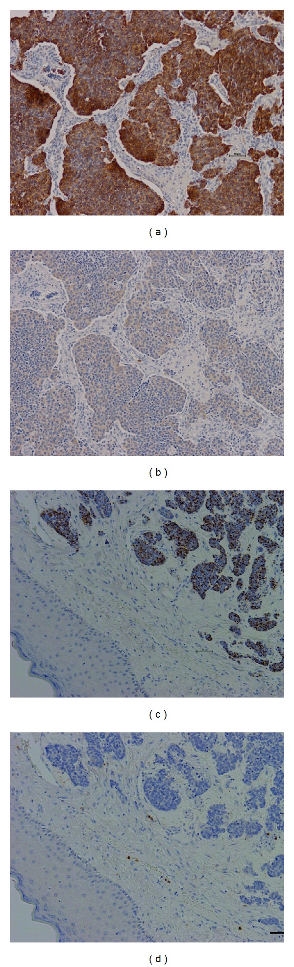
Due to the rarity of Merkel cell carcinoma (MCC), prospective clinical trials have not been practical. This study aimed to identify biomarkers with prognostic significance. While sixty-two patients were identified who were treated for MCC at our institution, only seventeen patients had adequate formalin-fixed paraffin-embedded archival tissue and followup to be included in the study. Patients were stratified into good, moderate, or poor prognosis. Laser capture microdissection was used to isolate tumor cells for subsequent RNA isolation and gene expression analysis with Affymetrix GeneChip Human Exon 1.0 ST arrays. Among the 191 genes demonstrating significant differential expression between prognostic groups, keratin 20 and neurofilament protein have previously been identified in studies of MCC and were significantly upregulated in tumors from patients with a poor prognosis. Immunohistochemistry further established that keratin 20 was overexpressed in the poor prognosis tumors. In addition, novel genes of interest such as phospholipase A2 group X, kinesin family member 3A, tumor protein D52, mucin 1, and KIT were upregulated in specimens from patients with poor prognosis. Our pilot study identified several gene expression differences which could be used in the future as prognostic biomarkers in MCC patients.


Figures

Figure 1
Figure 1
Representative example of histology of Merkel cell carcinoma from a tumor with poor prognosis. (a) Low power hematoxylin and eosin (H&E) stained photomicrograph. Arrows indicate regions of MCC to be microdissected. Dermis (De), epidermis (Ep), and sebaceous gland (SGI) are also indicated. (b) High power H&E image of same MCC. (c) Arcturus glass membrane hematoxylin counterstained serial section from same MCC. Areas to be laser capture microdissected are indicated by the arrow and red shading. The red punctate shading marks subsequent laser positioning. Using the ArcturusXT Microdissection System (molecular devices), these regions were transferred onto CapSure HS LCM Caps (Molecular Devices). Four caps were used per patient sample from the dissection of 1–4 sections representing approximately 5,000 tumor cells (1-2 mm2 area) captured per cap. (d) Image of the same membrane cut section after LCM. Magnification: (a) at 2X; (b), (c), and (d) at 10X. Scale bar = 50 μm.
Figure 2
Figure 2
Hierarchical clustering of Merkel cell carcinoma samples. Samples were clustered based upon the expression pattern of 45 differentially expressed genes between tumors of poor and good prognosis patients (P ≤ 0.01 and 1.5-fold cutoff). Individual samples are represented on the x-axis with poor prognosis patients in light blue (“poor”), good prognosis in green (“good”), and moderate prognosis in yellow (“mod”). Differentially expressed genes are shown on the y-axis. Clustering of (a) good and poor prognosis patients; (b) good, moderate, and poor prognosis patients. The scale of gene expression values are standardized such that genes are shifted to a mean of zero and scaled to a standard deviation of one.
Figure 3
Figure 3
Subnetwork of genes involved in regulating neoplasm metastasis, neurite outgrowth, and neurogenesis. Genes in red are upregulated in poor prognosis; blue, downregulated.
Figure 4
Figure 4
Immunohistochemistry in representative Merkel cell carcinoma tumors with good and poor prognoses. (a) intermediate KRT20 staining in poor prognosis tumor. (b) Weak KIF3A staining in poor prognosis tumor. (c) Weak punctate KRT20 staining in good prognosis tumor. (d) Negative (absent) KIF3A staining in good prognosis tumor. All images were taken at 10X magnification with Nikon digital DS-Fi1 high definition color camera on the Nikon Eclipse 90i. Scale bar = 50 μm.

References

    1. Toker C. Trabecular carcinoma of the skin. Archives of Dermatology. 1972;105(1):107–110. - PubMed
    1. Tang CK, Toker C. Trabecular carcinoma of the skin. An ultrastructural study. Cancer. 1978;42(5):2311–2321. - PubMed
    1. Hussain SK, Sundquist J, Hemminki K. Incidence trends of squamous cell and rare skin cancers in the swedish national cancer registry point to calendar year and age-dependent increases. Journal of Investigative Dermatology. 2010;130(5):1323–1328. - PubMed
    1. Lemos BD, Storer BE, Iyer JG, et al. Pathologic nodal evaluation improves prognostic accuracy in Merkel cell carcinoma: analysis of 5823 cases as the basis of the first consensus staging system. Journal of the American Academy of Dermatology. 2010;63(5):751–761. - PMC - PubMed
    1. Iyer JG, Koba S, Nghiem P. Toward better management of Merkel cell carcinoma using a consensus staging system, new diagnostic codes and a recently discovered virus. Actas Dermo-Sifiliograficas. 2009;100(supplement 2):49–54. - PMC - PubMed

LinkOut - more resources