. 2014 May;17(100):98-109.
doi: 10.1016/j.dnarep.2014.02.010. Epub 2014 Mar 11.

The spatial organization of non-homologous end joining: from bridging to end joining

Affiliations

The spatial organization of non-homologous end joining: from bridging to end joining

Takashi Ochi et al. DNA Repair (Amst). 2014 May.

Abstract

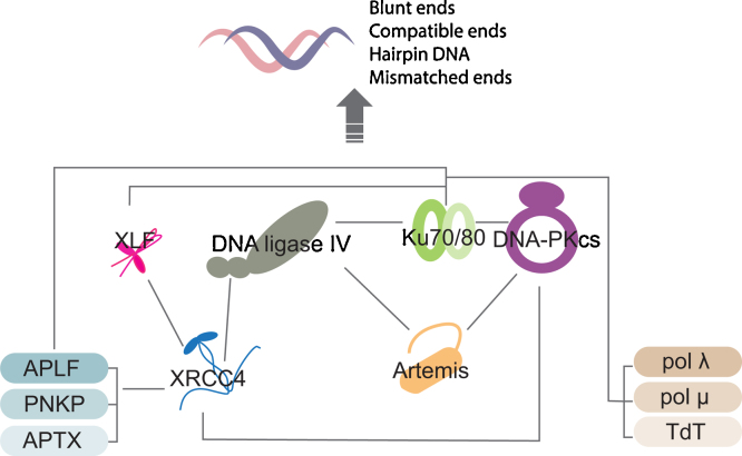
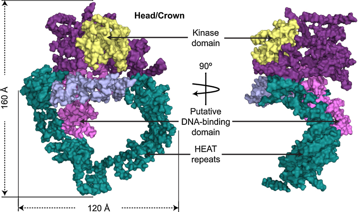
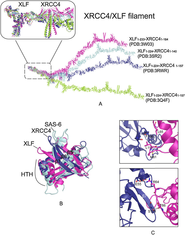
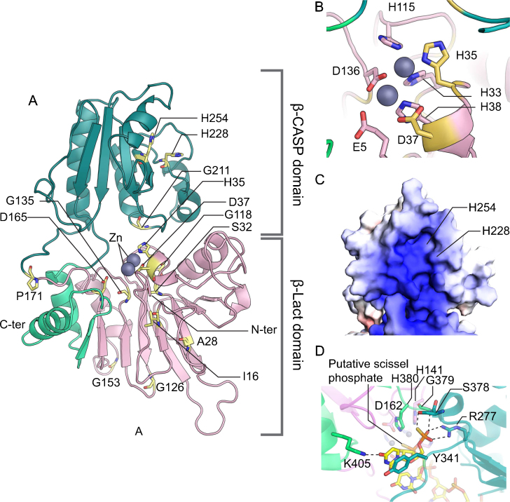
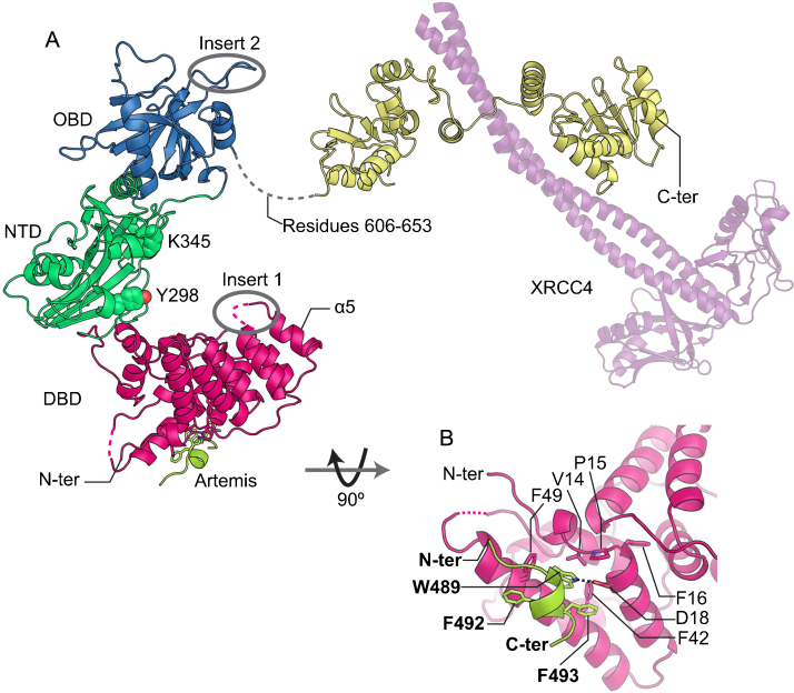
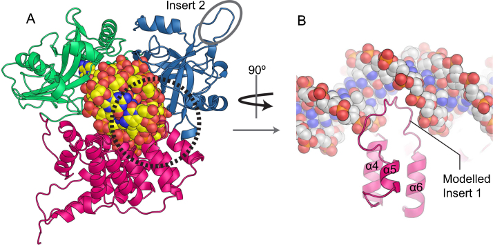
Non-homologous end joining (NHEJ) repairs DNA double-strand breaks generated by DNA damage and also those occurring in V(D)J recombination in immunoglobulin and T cell receptor production in the immune system. In NHEJ DNA-PKcs assembles with Ku heterodimer on the DNA ends at double-strand breaks, in order to bring the broken ends together and to assemble other proteins, including DNA ligase IV (LigIV), required for DNA repair. Here we focus on structural aspects of the interactions of LigIV with XRCC4, XLF, Artemis and DNA involved in the bridging and end-joining steps of NHEJ. We begin with a discussion of the role of XLF, which interacts with Ku and forms a hetero-filament with XRCC4; this likely forms a scaffold bridging the DNA ends. We then review the well-defined interaction of XRCC4 with LigIV, and discuss the possibility of this complex interrupting the filament formation, so positioning the ligase at the correct positions close to the broken ends. We also describe the interactions of LigIV with Artemis, the nuclease that prepares the ends for ligation and also interacts with DNA-PK. Lastly we review the likely affects of Mendelian mutations on these multiprotein assemblies and their impacts on the form of inherited disease.

Keywords: Artemis, DNA ligase IV; Cernunnus; DNA-PKcs; LIG4 syndrome; Non-homologous end joining; XLF; XRCC4.


Figures

Fig. 1
Fig. 1
Schematic representation of the NHEJ network. Grey lines indicate protein–protein interactions [64,80,88–90,161–170].
Fig. 2
Fig. 2
Molecular surface of the DNA-PKcs structure viewed perpendicular to the ring structure (left panel) and in the plane of the ring structure (right panel). The color code of the molecule is as follows: the ring structure is green; the forehead that is part of the ring structure is light purple; the putative DNA binding domain is pink; the larger C-terminal part is magenta, and the kinase domain is yellow (Adapted from ESRF Highlights, Newsletter, and Management Reports 2010 by BL Sibanda, D Chirgadze & TL Blundell).
Fig. 3
Fig. 3
The structure of the XRCC4/XLF complex. (A) XRCC4/XLF structures solved in four different groups [34–36,38]. One turn of XRCC4/XLF filament, which contains 6 copies of each XRCC4 and XLF molecules, is generated for comparison. Superimposition of the first XLF dimer molecules demonstrates varying curvatures of the filaments. (B) Superimposition of the head domains from XLF, XRCC4 and SAS-6. β6-7 and HTH are closer together in XLF than in XRCC4 and SAS-6. The PDB codes for structures here are 1IK9 (XRCC4), 2QM4 (XLF) and 2Y3V (SAS-6) . (C) The protein–protein interface of XRCC4/XLF, located in the head domain of each protein. The hydrophobic interface is shown in the top panel, while the bottom panel shows the polar interaction (indicated by grey dashed line). XLF is colored in red pink and XRCC4 is in deep purple. The XRCC4/XLF structure used is from .
Fig. 4
Fig. 4
Structural model of human Artemis. (A) Overall structural model of Artemis. β-Lact and β-CASP are shown in pink and emerald green, respectively. The C-terminal fold-back of β-Lact is highlighted with light green. Positions of residues, the missense mutations of which are found in patients carrying Artemis deficiency, are shown in stick representation with yellow color. Zinc atoms are shown in sphere representation. (B) The catalytic center of Artemis. The same color scheme as (A) is used here. (C) Surface representation of β-CAPS. The positions of H228 an H254 are indicated. (D) Close-up view of the catalytic center of an archaeal RNase (PDB code: 3IEM; unpublished). Dotted lines denote hydrogen bonds.
Fig. 5
Fig. 5
Structure of human LigIV in complex with a peptide corresponding residues 485–495 of Artemis. (A) Crystal structure of the LigIV/Artemis complex (PDB code: 3W1B; [82]) shown together with that of the LigIV/XRCC4 complex (PDB code: 3II6; [49]). DBD, NTD, OBD, BRCT domains and Artemis are shown in magenta, green, blue, yellow and lime, respectively. The XRCC4 dimer (purple) is presented in a transparent-cartoon representation. Dotted lines denote missing loops. Inset 1 and Inset 2 lies within the grey circles. Y298 and K345 are in sphere representation. (B) Details of the interaction between LigIV and Artemis. A hydrogen bond is indicated by a dotted line.
Fig. 6
Fig. 6
Structural model of LigIV bound nicked dsDNA. (A) Structural model of LigIV in complex with DNA. The model was built as described previously . The same color scheme as in Fig. 5A is used here. (B) Model of loop between α5 & α6 fitting into the major groove of DNA. The loop was modeled using RapperTK .
Fig. 7
Fig. 7
RS-SCID causing residues. (A) S32 and its surrounding residues. The same color scheme used in Fig. 5A is adopted here. (B) Residues around H228 and H254. Hydrogen bonds are indicated by dotted lines.
Fig. 8
Fig. 8
XLF mutants in Cernunnos-XLF deficiency patients. (A) R57, polar interaction is indicated by grey dashed line; (B) C123; (C) A25-R57 deletion. (D) R178X. Deletion protein sequences are shown in grey color.

References

    1. Lieber M.R. The mechanism of double-strand DNA break repair by the nonhomologous DNA end-joining pathway. Annu. Rev. Biochem. 2010;79:181–211. - PMC - PubMed
    1. Boulton S.J., Jackson S.P. Saccharomyces cerevisiae Ku70 potentiates illegitimate DNA double-strand break repair and serves as a barrier to error-prone DNA repair pathways. EMBO J. 1996;15:5093–5103. - PMC - PubMed
    1. Molly D.B.R., Bogue A., Chiyu Wang, Chengming Zhu V(D)J Recombination in Ku86-deficient mice: distinct effects on coding, signal, and hybrid joint formation. Immunity. 1997;7:37–47. - PubMed
    1. Kabotyanski E., Gomelsky L., Han J., Stamato T., Roth D. Double-strand break repair in Ku86- and XRCC4-deficient cells. Nucl. Acids Res. 1998;26:5333–5342. - PMC - PubMed
    1. Wang H., Perrault A.R., Takeda Y., Qin W., Wang H., Iliakis G. Biochemical evidence for Ku-independent backup pathways of NHEJ. Nucl. Acids Res. 2003;31:5377–5388. - PMC - PubMed

Publication types

MeSH terms

LinkOut - more resources